Membranous wall of trachea (Latin: paries membranaceus tracheae)
Also called the posterior membranous wall
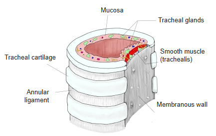
· The tracheal cartilages are open at the back, and the membranous wall closes the gap between their ends, so the trachea is flat behind. Its main part is the trachealis, which joins the ends of the cartilages; large tracheal glands lie in and outside it.
· Not to be confused with the membranous wall of the tympanic cavity (the eardrum wall of the middle ear), a different structure with the same short name.
of the
trachea
- Tracheal cartilages : 15–20 C-shaped bars of hyaline cartilage, each about 3–4 mm wide.
- Annular ligaments : continuations of the connective tissue membrane (fibroelastic membrane) that wraps the whole set of cartilages.
- Mucosa : well developed and lining the lumen; it has no folds and contains the tracheal glands, blood vessels and nerves.
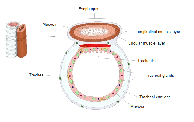
- Membranous wall : the back wall facing the esophagus, made mainly of smooth muscle (the trachealis), with large tracheal glands.
· Because the membranous wall is soft, the esophagus behind it can bulge forward into it when a bolus of food passes down; the wall is thought to make this widening of the esophagus easier.
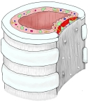 Trachea (cross-section)
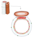 Trachea and esophagus (cross-section)
Reference websites
[Wikipedia]
· Trachea, Wikipedia.
[Websites with illustrations]
· Trachea in cross-section: tracheal cartilage, trachealis muscle and esophagus (Elsevier (journal figure))

· Smooth muscle in the tracheal wall (Fine Art America (Asklepios Medical Atlas))

· Posterior membranous trachea and esophagus (Cambridge University Press)

Related terms
References and sources
- Further reading: Trachea, Wikipedia.
- Federative International Programme for Anatomical Terminology (FIPAT). Terminologia Anatomica, 2nd ed. (TA2), 2019.