Esophagus (Latin: oesophagus)
- Overview
- Parts (cervical, thoracic, abdominal)
- Physiological constrictions
- Structure (mucosa, submucosa, muscle layer, adventitia)
- Glands
- Vessels and nerves
- Development
- References
Overview
- Respiratory system
- Digestive system : oral cavity, pharynx, esophagus, stomach, small intestine, large intestine
- Urinary system
- Genital system
- Peritoneum
- Endocrine glands
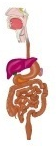
· For reference: from the incisor teeth to the cardia of the stomach is about 37–40 cm, and from the incisor teeth to the beginning of the esophagus about 15 cm.
· It is a tube about 25 cm long, 2–3 cm wide, with a wall about 4 mm thick.
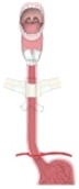 Oral cavity to cardia
(frontal section)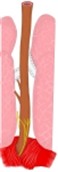 Beginning to esophageal hiatus
(posterior view)
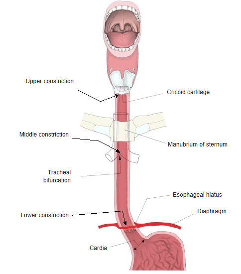
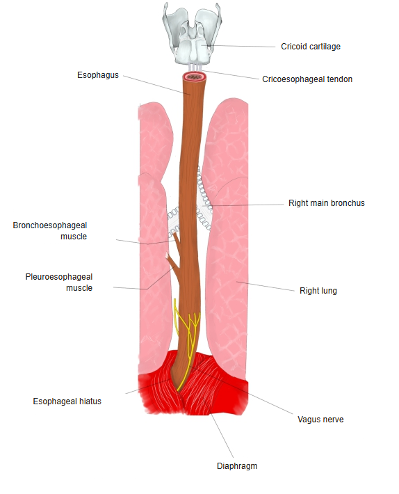
Parts
According to its level, the esophagus is divided into three parts: cervical, thoracic and abdominal.
esophagus
- Cervical part : above the upper border of the manubrium of the sternum; continues from the pharynx. Length about 4–5 cm.
- Thoracic part : the longest part, from the upper border of the manubrium to the esophageal hiatus of the diaphragm. Length about 18–20 cm.
- Abdominal part : the shortest part, from the esophageal hiatus to the cardia of the stomach. Length about 1–2 cm.
· The lengths are approximate and vary between individuals and sources.
[Cervical part]
· The short part above the level of the upper border of the manubrium of the sternum.
· It descends straight between the trachea and the vertebral column, inclining a little to the left in its lower part.
· It is joined to the trachea by loose connective tissue; the right and left recurrent laryngeal nerves run in this tissue or a little lateral to it.
· The right and left common carotid arteries run lateral to the cervical part.
[Thoracic part]
· After entering the thoracic cavity it runs more and more to the left of the trachea, and a little below the tracheal bifurcation it passes almost directly behind the left main bronchus.
· It descends in front of the thoracic vertebrae and the thoracic aorta (at first to the right of the aorta, then gradually in front of it), and passes through the esophageal hiatus of the diaphragm into the abdominal cavity; where it pierces the diaphragm it lies in front of and to the left of the aorta.
· In its upper part the azygos vein runs upwards on its right side and the thoracic duct behind it.
[Abdominal part]
· After entering the abdominal cavity it bends a little to the left (sometimes upwards) and reaches the cardia of the stomach in front of and to the left of the 11th thoracic vertebra.
· It is the shortest of the three parts.
Physiological constrictions
the esophagus
- Upper constriction : at the beginning of the esophagus, at the level of the 6th cervical vertebra; about 15 cm from the incisor teeth.
- Middle constriction : near the tracheal bifurcation, where the esophagus is crossed by the aortic arch and the left main bronchus (4th–5th thoracic vertebrae); about 25 cm from the incisor teeth.
- Lower constriction : where it passes through the esophageal hiatus of the diaphragm (about the 10th–11th thoracic vertebrae); about 40 cm from the incisor teeth.
At these constrictions the esophagus cannot widen easily because of its surroundings, so swallowed material passes with difficulty and tends to lodge there.
- Constrictions
Structure
the esophagus
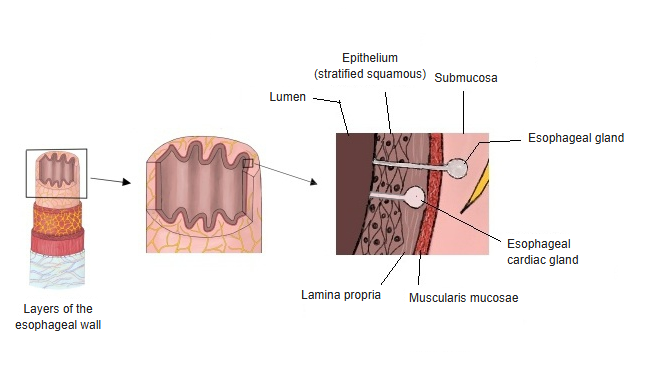
- Mucosa
- Epithelium : stratified squamous epithelium with Langerhans cells.
- Lamina propria : dense connective tissue with elastic fibres; contains the esophageal cardiac glands (not always present).
- Muscularis mucosae : smooth muscle fibres; the best developed in the digestive tract, especially in the lower part.
- Submucosa : loose connective tissue that forms the folds; contains the esophageal glands (mixed glands). The submucosal plexus (Meissner's plexus) is weak and hard to see.
- Muscle layer
- Inner circular layer : C-shaped bundles arranged in a discontinuous spiral.
- Outer longitudinal layer : strongly differentiated; sends bundles of fibres to neighbouring structures.
- Adventitia : a thick fibrous layer that binds the esophagus to neighbouring organs and carries fairly large nerves and vessels.
The description of each layer follows.
[Mucosa]
Epithelium
· Made of stratified squamous epithelium.
· It is generally not keratinized, but slight keratinization is seen in places.
· Among the epithelial cells are Langerhans cells, which take part in immune defence.
· Its junction with the epithelium of the stomach is a saw-toothed (zig-zag) line.
Lamina propria
· A dense connective tissue layer containing elastic fibres, with the esophageal cardiac glands.
· It forms small papillae projecting into the epithelium.
· (Lower) esophageal cardiac glands: not always present; mucous glands confined to the lowest part of the esophagus.
· Upper esophageal cardiac glands: structurally the same as the lower ones; found between the upper end of the esophagus and the level of the 5th tracheal cartilage in about 70% of adults and about 90% of fetuses.
Muscularis mucosae
· Made of smooth muscle fibres running along the long axis of the esophagus.
· It is the best developed muscularis mucosae of the digestive tract, and is stronger in the lower part of the esophagus than in the upper part.
· Above, it continues into the fibrous membrane of the pharynx; below, into the muscularis mucosae of the stomach.
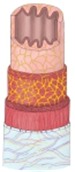 Structure of the esophagus
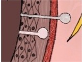 Mucosa of the esophagus
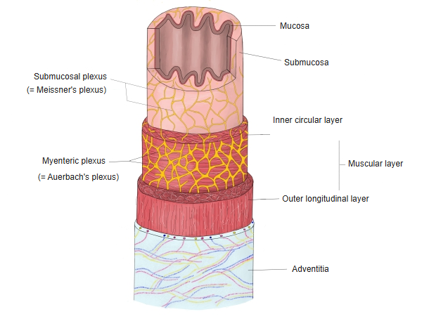
[Submucosa]
· Loose connective tissue that forms the folds. It contains the esophageal glands (mixed glands in which mucous cells predominate), but how well they are developed varies greatly between individuals.
· The ducts of the glands often widen along their course, and groups of lymphocytes are often found around them.
· The submucosal plexus (Meissner's plexus) is poorly developed and hard to see.
[Muscle layer]
The upper third of the esophagus is made of striated muscle, the middle third of striated and smooth muscle, and the lower third entirely of smooth muscle. The layer is divided into an inner circular and an outer longitudinal layer.
Inner circular layer
· It is not a continuous spiral; C-shaped interrupted bundles are arranged in a spiral.
· The outer longitudinal layer runs at right angles to the inner circular layer.
· Swallowed food is carried along by peristalsis of the muscle layer.
Outer longitudinal layer
· Compared with the inner circular layer it is strongly differentiated, and some of its fibres pass to neighbouring structures:
1. Cricoesophageal tendon: the tendon by which the longitudinal muscle of the esophagus is attached in front to the back of the lamina of the cricoid cartilage.
2. Bronchoesophageal muscle: a slip of smooth muscle passing between the esophagus and the left main bronchus.
3. Pleuroesophageal muscle: a slip of smooth muscle passing between the esophagus and the left mediastinal pleura.
· Between the inner circular and outer longitudinal layers lies the well-developed myenteric plexus (Auerbach's plexus), which contains nerve cells.
· In the upper part, where the muscle is striated, this plexus is poorly developed.
- Esophagus (posterior view)
[Adventitia]
A thick fibrous layer that binds the esophagus to neighbouring organs or to the body wall; it carries fairly large nerves and blood vessels.
Glands
esophagus
- Esophageal cardiac glands : site: the mucosa (especially near the junction with the stomach at the lower end, but also at the upper end of the esophagus).
- Esophageal glands : site: the submucosa. Mixed glands in which mucous cells predominate. Also called esophageal submucosal glands or proper esophageal glands.
Esophageal cardiac glands
Mucous glands in the lamina propria of the mucosa near the cardiac orifice (the junction of the esophagus and stomach). They secrete neutral mucin, which protects the esophagus from acid gastric juice. They are simple tubular or branched tubular glands.
Esophageal glands
The esophageal submucosal glands are compound tubuloalveolar glands with some serous cells. They are more numerous in the upper third of the esophagus and secrete acid mucin, which lubricates the passage of food.
- Structure of the esophagus
- Mucosa of the esophagus
Vessels and nerves
[Arteries]
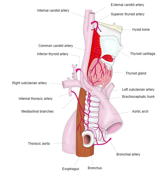
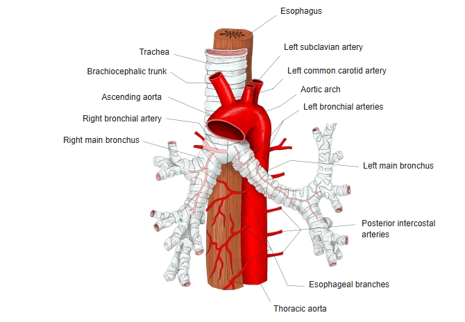
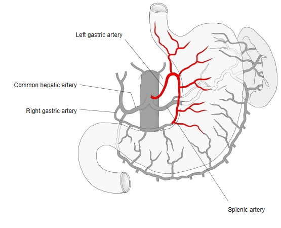
The upper part is supplied by the esophageal branches of the inferior thyroid artery, the middle part by the esophageal arteries (branches of the thoracic aorta), and the lower part by the esophageal branches of the left gastric artery.
 Inferior thyroid artery
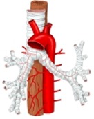 Esophageal arteries
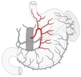 Left gastric artery
[Veins]
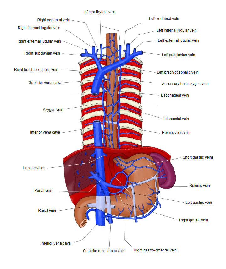
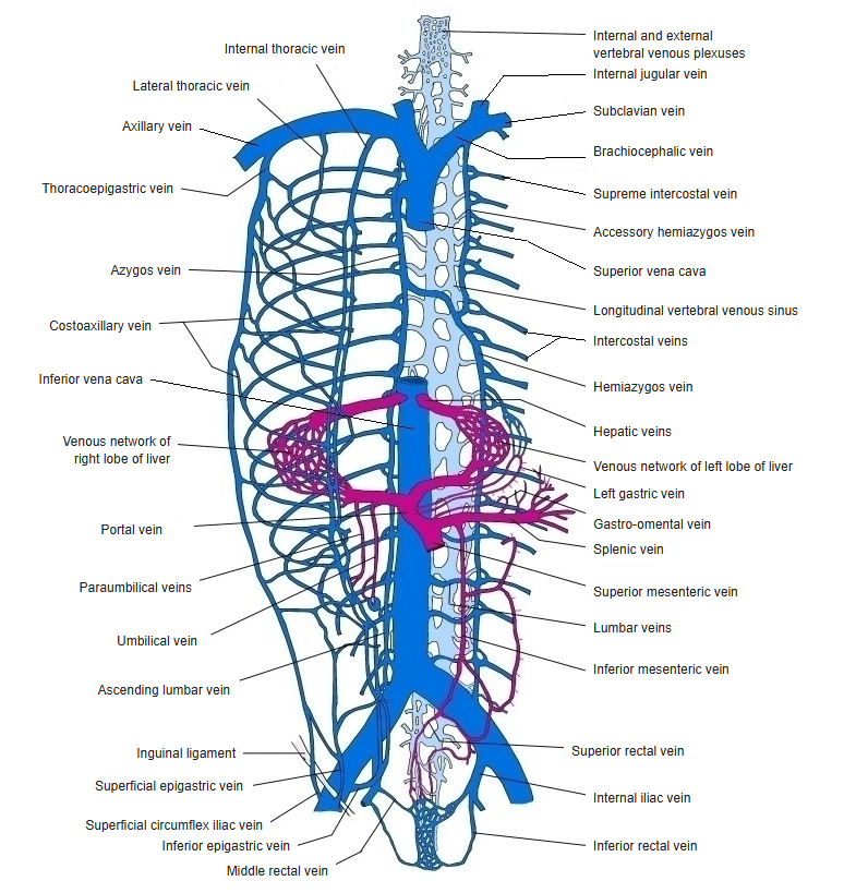
The esophageal veins drain into the inferior thyroid veins, the azygos vein and the hemiazygos vein (connected through the azygos vein with the superior vena cava), and into the left gastric (coronary) vein, through which they communicate with the portal vein.
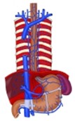 Veins of the esophagus
and stomach Azygos and hemiazygos
veins
[Lymphatics]
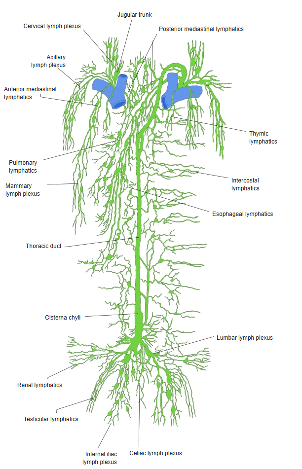
Lymph from the cervical part drains to the deep and superficial cervical lymph nodes, and lymph from the thoracic part to the posterior mediastinal lymph nodes.
 Lymph vessels of the trunk
[Nerves]
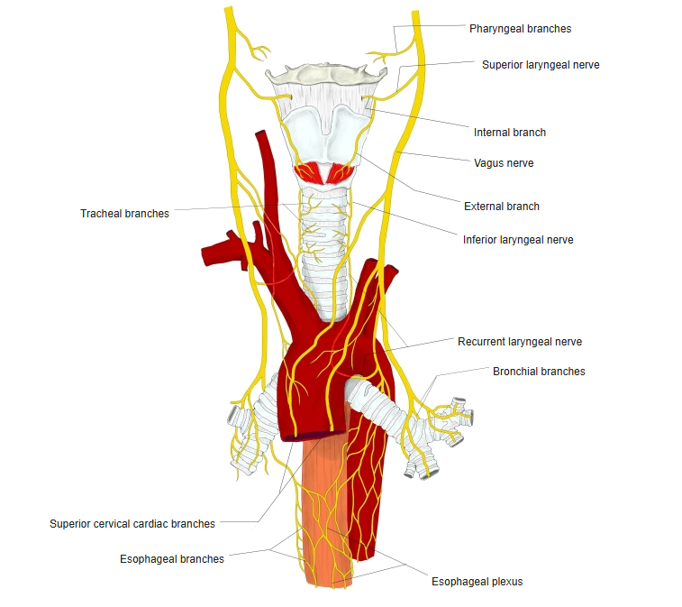
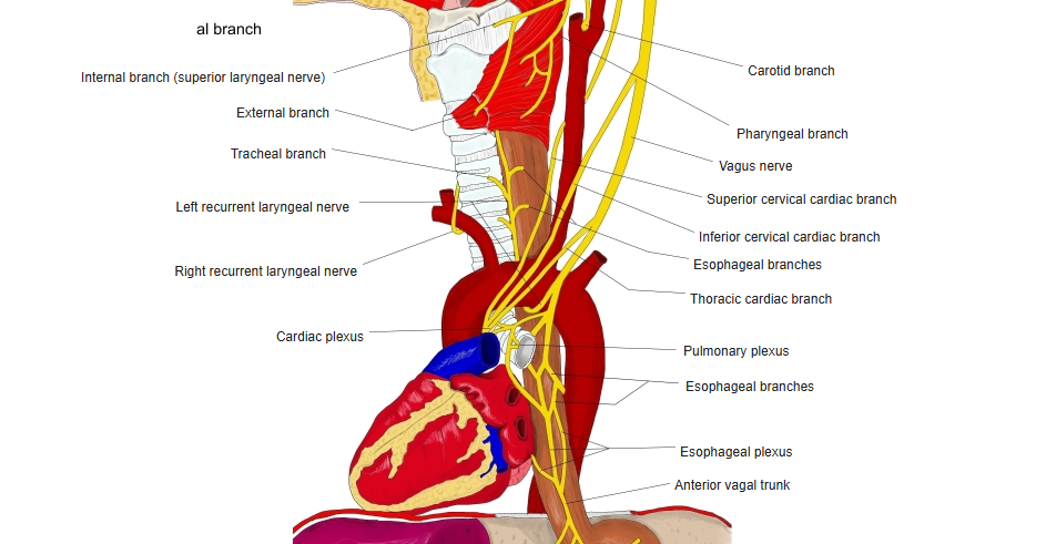
Branches of the anterior and posterior esophageal plexuses, formed by the vagus nerves and the sympathetic trunks. Sensory fibres come from the 5th–8th thoracic nerves.
 Vagus nerve
 Vagus nerve
Development
The esophagus develops from part of the foregut. Early in development it is fairly short and wide and compressed from side to side. At the beginning of the 2nd month of fetal life it suddenly lengthens, although its diameter grows little. At birth it is about 8–10 cm long, and by about 3 years of age its length has roughly tripled.
References and sources
The esophagus is a fibromuscular tube, about 25 cm long in adults, through which food passes by peristalsis from the pharynx to the stomach. It runs behind the trachea and the heart, passes through the diaphragm and opens into the uppermost part of the stomach. During swallowing the epiglottis tilts back so that food does not enter the larynx.
From the lumen outwards its wall consists of the mucosa, the submucosa, the muscle layers and an outer connective tissue layer. The mucosa is a non-keratinized stratified squamous epithelium, in contrast to the single layer of columnar cells of the stomach; the change between them is seen as a zig-zag line. Striated muscle predominates in the upper third and smooth muscle elsewhere. There is a muscular sphincter at each end; the lower one helps to prevent reflux of acid stomach contents. The smooth muscle is supplied by autonomic nerves (sympathetic fibres from the sympathetic trunk and parasympathetic fibres in the vagus nerve), and the striated muscle by motor fibres carried in the vagus nerve.
The esophagus may be affected by reflux of gastric contents, cancer, bleeding varices (dilated veins), tears, narrowing and disorders of movement, which can cause difficult or painful swallowing or chest pain.
- Further reading: Esophagus, Wikipedia.