Myofibril (Latin: myofibrilla)
Also called the muscle fibril
A myofibril is
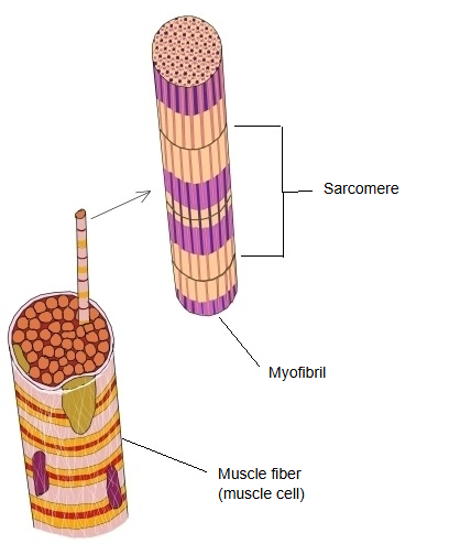
 Muscle fiber, myofibril and sarcomere
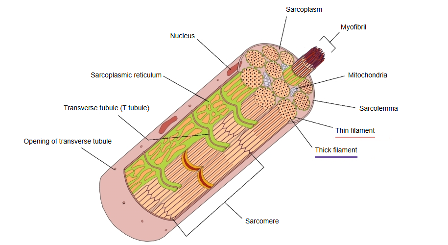
 Skeletal muscle fiber
Below is a simple diagram of the levels of structure of a muscle.
From myofilament to muscle
Reference websites
[Wikipedia]
· Myofibril, Wikipedia.
Bands
Along its length a myofibril shows two kinds of band, which alternate. Each myofibril is a chain of sarcomeres placed end to end; the bands of neighbouring myofibrils line up across the fibre, which gives striated muscle its striped look.
| 1 | A band (dark band) | Looks dark under the light and electron microscope. It is the part of each sarcomere that contains the thick (myosin) filaments, and its length stays the same during contraction. Its pale centre is the H zone, crossed by the M line. | 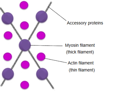 |
| 2 | I band (light band) | Looks light. It is the part on each side of a Z disc that contains only thin (actin) filaments; it becomes shorter during contraction as the thin filaments slide in between the thick filaments. |
In cross-section, where the filaments overlap, each thick filament is surrounded by thin filaments in a regular hexagonal pattern. Each myofibril is wrapped by the sarcoplasmic reticulum, with transverse tubules and mitochondria lying between the myofibrils. Muscle contracts when the thin filaments slide along the thick filaments, so every sarcomere, and with it the whole myofibril, shortens.
Related terms
References and sources
- Further reading: Myofibril, Wikipedia.
- Federative International Programme for Anatomical Terminology (FIPAT). Terminologia Anatomica, 2nd ed. (TA2), 2019.