Outer phalangeal cell
Also called Deiters' cell; cell of Deiters
= Cells of Deiters
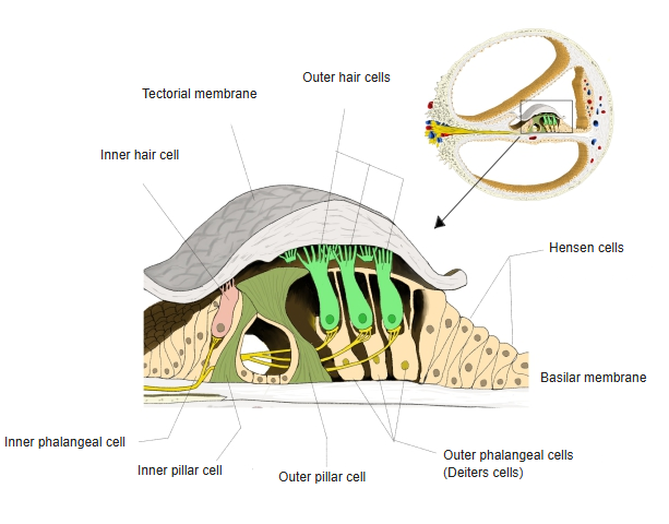
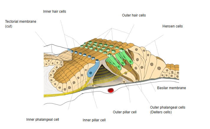
An outer phalangeal cell is a supporting cell of the spiral organ (organ of Corti) that holds an outer hair cell in a cup on its upper surface. The cells stand on the basilar membrane in rows outside the outer pillar cells, usually three rows (sources differ; more rows are found towards the apex of the cochlea). From each cell a thin phalangeal process rises between the hair cells to the surface, where the heads of the processes join the tops of the hair cells to form the reticular membrane (reticular lamina). A bundle of microtubules and microfilaments runs through each cell from base to apex and stiffens it. The space between the outer pillar cells, the outer hair cells and these cells is the space of Nuel; the tunnel of Corti lies inside the outer pillar cells. The cells are named after the anatomist Otto Deiters.
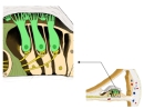 Outer phalangeal cells (green)
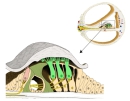 Spiral organ (section)
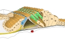 Spiral organ (3D view)
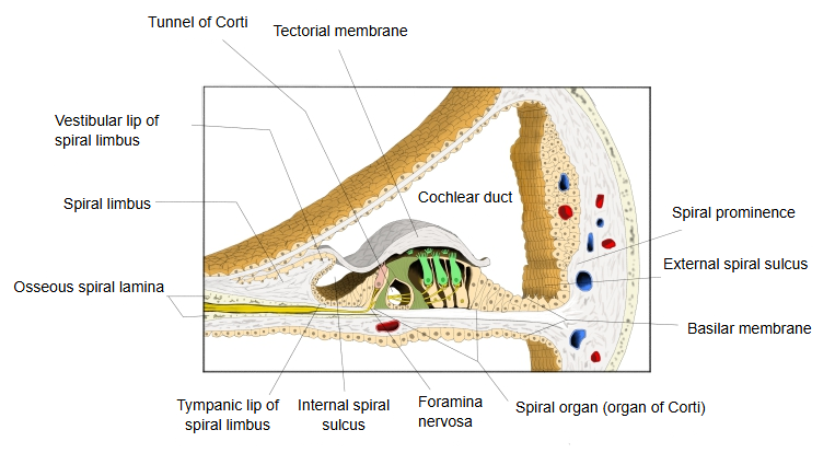
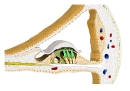 Cochlear duct (section)
Related terms
- Organ of Corti
- Spiral organ (organ of Corti)
- Basilar membrane
- Reticular membrane
- Tunnel of Corti
- Phalangeal process
- Cochlear duct
References and sources
- Further reading: Deiters cell, Wikipedia.
- Federative International Programme for Anatomical Terminology (FIPAT). Terminologia Anatomica, 2nd ed. (TA2), 2019.