Proximal epiphysis of radius (Latin: epiphysis proximalis radii)
Also called the upper end of radius
· It is also called the upper end of the radius.
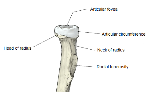
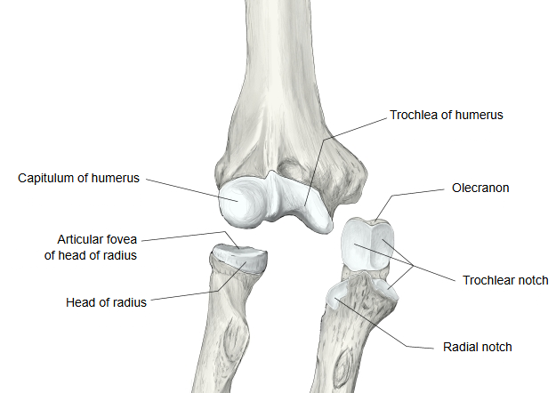
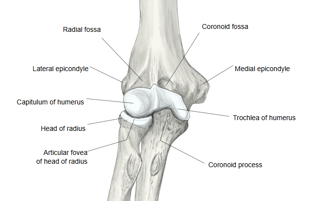
· The shallow hollow, the articular fovea, articulates with the capitulum of the humerus ⇒ humeroradial joint (one of the joints of the elbow joint).
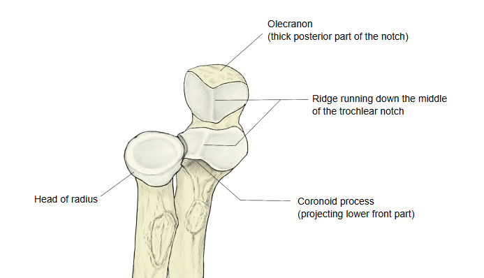
· The articular circumference articulates with the radial notch of the ulna ⇒ proximal radioulnar joint (one of the joints of the elbow joint); the annular ligament of radius holds the head in place.
· Anatomy textbooks describe these parts of the upper end: the head of radius, the articular fovea, the articular circumference and the neck of radius. Below the neck lies the radial tuberosity. Sources differ on how far the term reaches: in the strict developmental sense the proximal epiphysis is only the head, which ossifies from its own centre; descriptions of the upper end usually include the neck and the radial tuberosity.
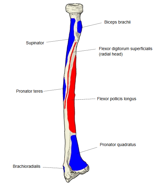
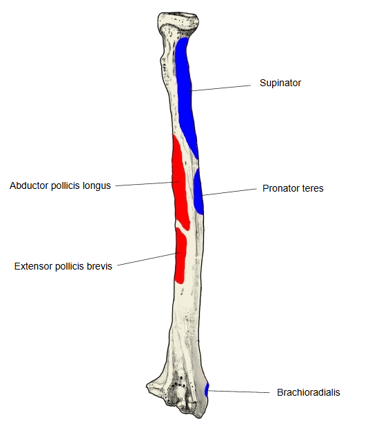
· Muscles attached: insertion of supinator (a small part, around the neck). Biceps brachii is inserted into the radial tuberosity just below.
· Ligament attached: the quadrate ligament, which joins the neck to the ulna. The radial collateral ligament of the elbow is attached to the annular ligament, not to the radius itself.
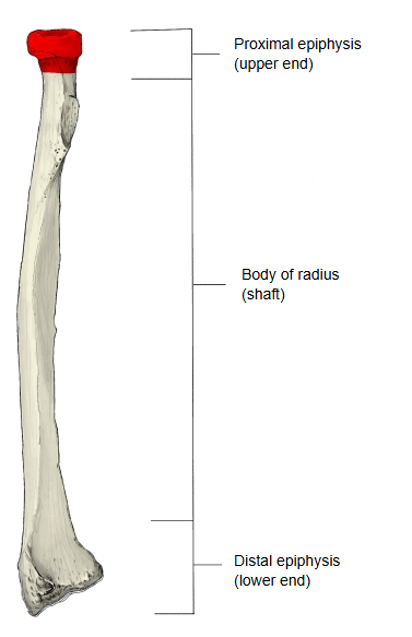
- Proximal epiphysis : the disc-shaped enlargement with a shallow hollow = upper end.
- Body of radius : the three-sided middle part that forms the shaft; it has three borders and three surfaces.
- Distal epiphysis : much wider from side to side than the proximal epiphysis = lower end.
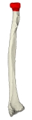 Right radius (anterior)
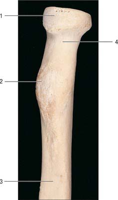
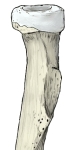 Upper end of right radius
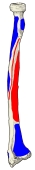 Muscle attachments (anterior)
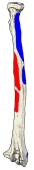 Muscle attachments (posterior)
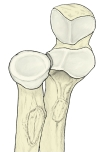 Upper ends of radius and ulna
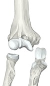 Right elbow (bones separated)
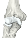 Right elbow (anterior)
Sites with illustrations
· Upper end of the radius (photograph, Clinical Gate)

Where it is found
Related terms
- Head of radius
- Neck of radius
- Radial tuberosity
- Articular fovea of radius
- Articular circumference of radius
- Distal epiphysis of radius
- Proximal radioulnar joint
References and sources
- Further reading: Radius (bone), Wikipedia.
- Further reading: Head of radius, Wikipedia.
- Federative International Programme for Anatomical Terminology (FIPAT). Terminologia Anatomica, 2nd ed. (TA2), 2019.