Semilunar line (Latin: linea semilunaris)
Also called Spigelian line
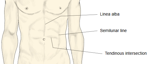
· It shows more clearly in people with well-developed abdominal muscles and little body fat, as a shallow curved groove at each side of rectus abdominis.
rectus
abdominis
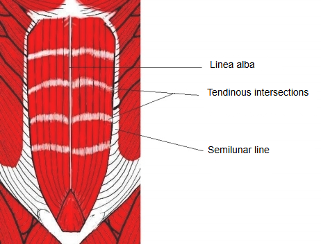
- Linea alba : a single line of connective tissue running vertically down the middle (the midline) between the right and left rectus abdominis
- Tendinous intersections : 3–4 bands of connective tissue that cross the belly of rectus abdominis and divide it into segments
- Semilunar line : 2 lines, one on each side, curving along the lateral edge of rectus abdominis
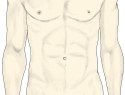 Surface of the abdomen
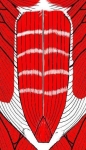 Rectus abdominis
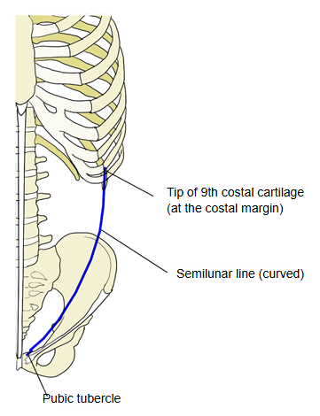
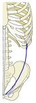 Course of the semilunar line
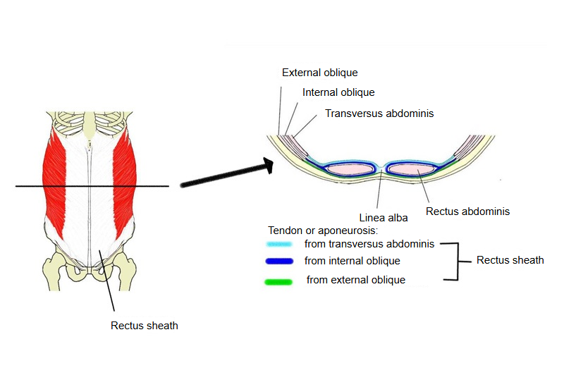
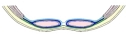 Rectus sheath (cross-section)
Structure
The semilunar line (Spigelian line) is a curved line, one on each side of the front abdominal wall, that follows the lateral border of rectus abdominis from the tip of the 9th costal cartilage down to the pubic tubercle. Along it the aponeurosis of the internal oblique splits to enclose rectus abdominis; it is joined in front by the aponeurosis of the external oblique and, above the arcuate line, behind by the aponeurosis of transversus abdominis. Together these layers form the rectus sheath. A hernia through the weak area beside the line is a Spigelian hernia.
Reference websites
[Wikipedia]
· Linea semilunaris, Wikipedia.
· Rectus sheath, Wikipedia.
[Websites with illustrations]
· Rectus sheath above the arcuate line (cross-section), with the linea semilunaris (Radiopaedia)

Related terms
- Rectus abdominis
- Internal oblique
- External oblique
- Transversus abdominis
- Pubic tubercle
- Inguinal ligament
References and sources
- Further reading: Linea semilunaris, Wikipedia.
- Further reading: Rectus sheath, Wikipedia.
- Federative International Programme for Anatomical Terminology (FIPAT). Terminologia Anatomica, 2nd ed. (TA2), 2019.