Tarsal plates (Latin: tarsi palpebrarum)
Also called the tarsi (of the eyelids) or the tarsus
· Anatomy texts give its length as about 20 mm and its greatest thickness, at the middle, as about 0.7 mm; most English sources give about 25-30 mm and about 1 mm (sources differ).
The table below summarises the differences between the upper and lower tarsi.
| Superior tarsus | Inferior tarsus | |
|---|---|---|
| Height | about 10 mm | about 5 mm |
| Shape | crescent | elliptical |
| Number of tarsal glands | about 25 (30-40) | about 20 (20-30) |
| Other | joined by the superior tarsal muscle | joined by the inferior tarsal muscle |
The number of tarsal glands differs considerably between sources; figures of 30-40 in the upper and 20-30 in the lower lid are also given.
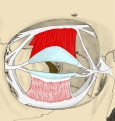 Tarsi (and tarsal muscles) and ligaments
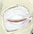 Tarsi and orbital septum
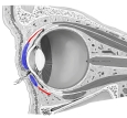 Tarsi (and tarsal muscles)
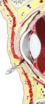 Front of the orbit (sagittal section)
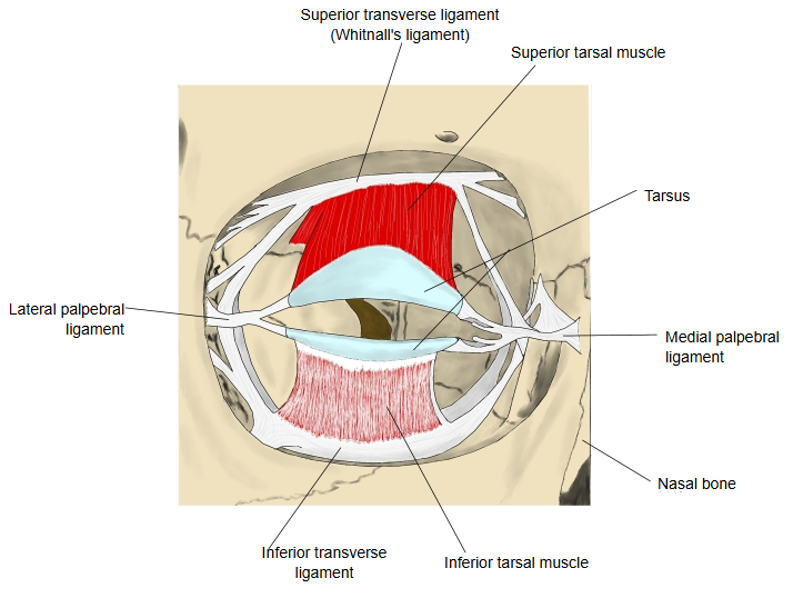
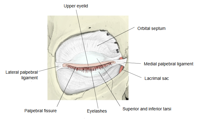
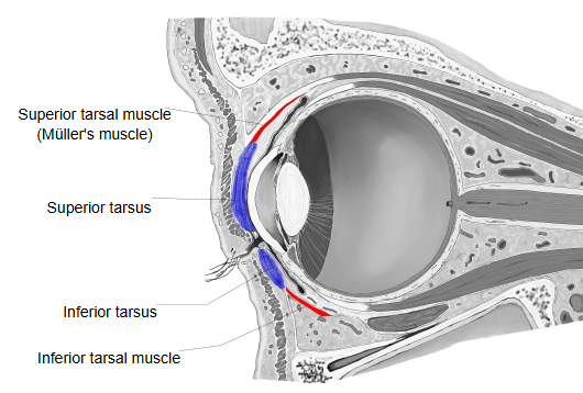
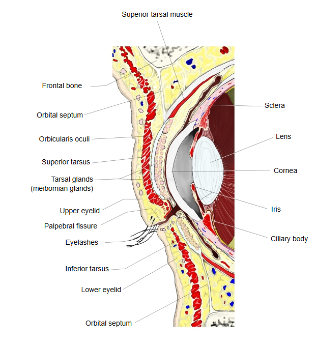
Each tarsus is tallest in the middle of the eyelid and narrows towards both ends. It is made of firmly interwoven bundles of connective tissue, like felt. At the medial angle of the eye the medial palpebral ligament blends with the tarsi; it runs from there to the frontal process of the maxilla, just under the skin in front of the lacrimal sac, and its front edge can be felt when the eye is closed. The lateral palpebral ligament lies behind the orbicularis oculi and the orbital septum and attaches to the zygomatic bone 2-3 mm behind the orbital margin.
Reference websites
[Wikipedia]
· Tarsus (eyelids), Wikipedia.
[Websites with illustrations]
· Eyelid anatomy (illustration) (Clinical Gate)
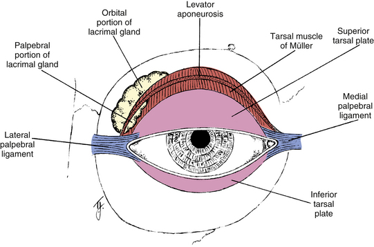
Superior tarsus
The superior tarsus is the larger: crescent-shaped, about 10 mm high in the middle and narrowing towards its ends. The smooth superior tarsal muscle is attached to its upper border, and fibres of the aponeurosis of levator palpebrae superioris attach to its front surface.
Inferior tarsus
The inferior tarsus is smaller and thinner, elliptical, about 5 mm high. The free (ciliary) margins of both plates are thick and straight.
Relations
The attached (orbital) margins are joined to the rim of the orbit by the orbital septum. The lateral ends are attached to the zygomatic bone by the lateral palpebral ligament (with the lateral palpebral raphe in front), and the medial ends to the frontal process of the maxilla by the medial palpebral ligament. Along each tarsus runs a vertical row of tarsal (meibomian) glands, modified sebaceous glands whose lipid secretion slows the evaporation of tears and keeps the eye moist.
Related terms
References and sources
- Further reading: Tarsus (eyelids), Wikipedia.
- Federative International Programme for Anatomical Terminology (FIPAT). Terminologia Anatomica, 2nd ed. (TA2), 2019.