Orbital septum (Latin: septum orbitale)
· It can be divided into the superior orbital septum and the inferior orbital septum.
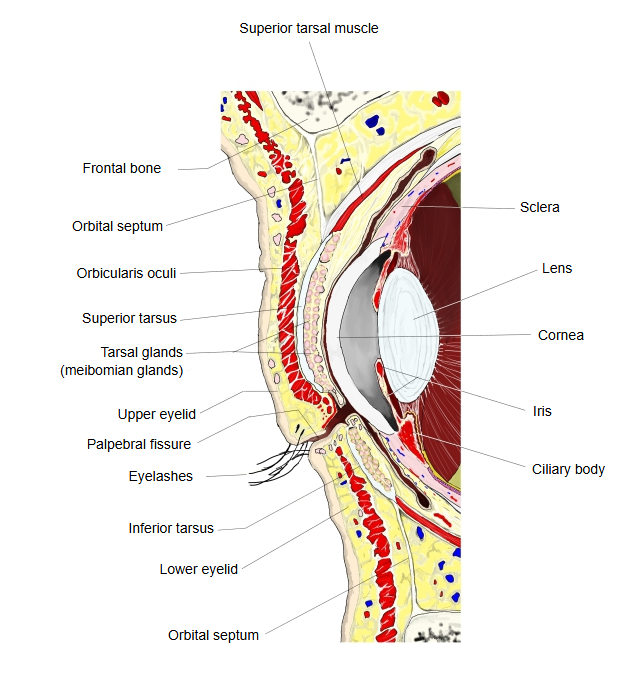
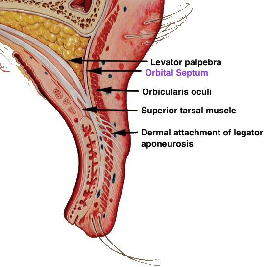
1. 1. Superior orbital septum: runs down from the orbital margin and attaches near the insertion of the levator palpebrae superioris, blending with its aponeurosis above the superior tarsus.
2. 2. Inferior orbital septum: runs up from the orbital margin and attaches to the lower edge of the inferior tarsus.
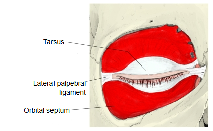
· At the corners of the eye the upper and lower septa meet the medial palpebral ligament and the lateral palpebral ligament; together with the orbicularis oculi they support the eyelids.
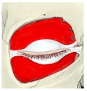 Left orbit (anterior part)
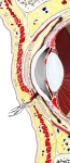 Eyelids and front of orbit (sagittal section)
· In more detail:
The orbital septum is a sheet of connective tissue that joins the orbital margin to the tarsi. It divides each eyelid into an anterior and a posterior layer: it lies behind the orbicularis oculi and in front of the orbital fat body. In the upper eyelid it blends, deep to the orbicularis oculi, with the aponeurosis of the levator palpebrae superioris. When the eyes are closed, the septum and the tarsi together cover the whole orbital opening. Medially it is thin and passes behind the medial palpebral ligament to the posterior lacrimal crest. Vessels and nerves passing from the orbit to the face and scalp pierce it.
Reference websites
[Wikipedia]
· Orbital septum, Wikipedia.
[Websites with illustrations]
· Orbital septum separating preseptal and postseptal (orbital) cellulitis (figure) (RCNI)

· Section of the upper eyelid with the orbital septum (figure) (Pediatric EM Morsels)
Related terms
- Superior orbital septum
- Inferior orbital septum
- Eyelids
- Orbital fat body
- Levator palpebrae superioris
- Orbicularis oculi
- Lateral palpebral ligament
References and sources
- Further reading: Orbital septum, Wikipedia.
- Federative International Programme for Anatomical Terminology (FIPAT). Terminologia Anatomica, 2nd ed. (TA2), 2019.