Zygapophysial joints (Latin: articulationes zygapophysiales)
Also called Facet joints
What are the zygapophysial joints?
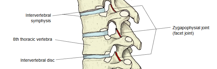
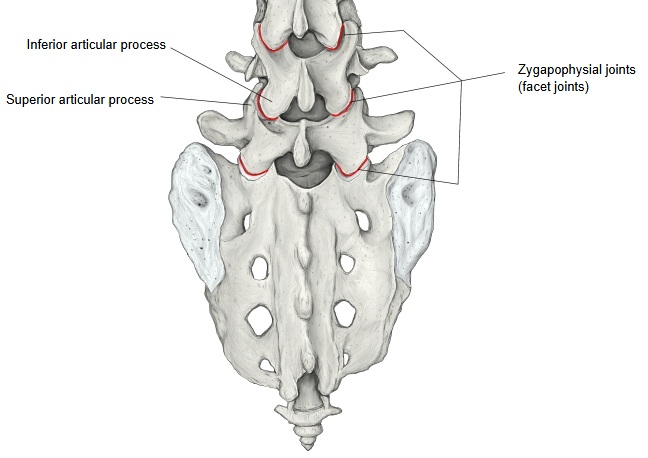
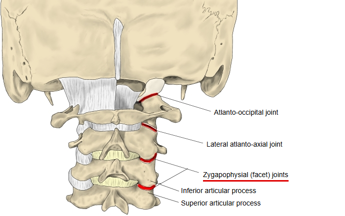
Zygapophysial joint: inferior articular process (inferior articular facet) of the upper vertebra ⇔ superior articular process (superior articular facet) of the lower vertebra
- Highest: between the inferior articular process of the 2nd cervical vertebra (axis) and the superior articular process of the 3rd cervical vertebra. The joint between the 1st cervical vertebra (atlas) and the axis at the same position is called the lateral atlanto-axial joint.
- Lowest: between the inferior articular process of the 5th lumbar vertebra and the superior articular process of the sacrum. Note that some illustrations do not count this joint (part of the lumbosacral joint) as a zygapophysial joint.
- The joint capsule is thin but strong; its front (inner) and back (outer) sides are reinforced by strong ligaments.
Articular surfaces
| Region | Orientation of the articular surfaces |
|---|---|
| Cervical vertebrae | Slope from front and above to back and below. |
| Thoracic vertebrae | Close to the frontal (coronal) plane; the right and left surfaces lie on an arc centred on the front of the vertebral body. |
| Lumbar vertebrae | Almost sagittal, but curved medially so that their front parts face nearer the frontal plane. |
Bones
More about the joints
Each zygapophysial joint is a plane joint between the inferior articular process of the upper vertebra and the superior articular process of the lower one. The joints are loose in the cervical region and tight in the thoracic region. The capsule is thin but strong. On its front (inner) side it is reinforced by the lateral part of the ligamentum flavum, which continues over the joint, so that the two together extend to the back margin of the intervertebral foramen; its back (outer) side is also reinforced by strong ligament fibres.
The joints are synovial plane joints, two in each motion segment of the spine (a pair of adjacent vertebrae with the tissues between them). Each is supplied by the medial branches of the dorsal rami of the spinal nerves. Their mechanical role is to guide and limit the movement of the segment. In the lumbar region they protect the segment from forward shearing forces, excessive rotation and excessive flexion; their surfaces allow flexion and extension but limit rotation, and they have little effect on side bending. In the thoracic region they limit flexion and forward sliding of the vertebrae and allow rotation. Their function can be disturbed by degeneration, dislocation, fracture, instability after injury, osteoarthritis and surgery. The popping sound heard during spinal manipulation ("cracking the back") comes from gas bubbles forming in the synovial fluid of these joints.