Latin: musculus adductor brevis
Overview
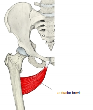
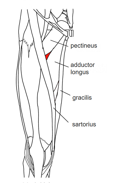
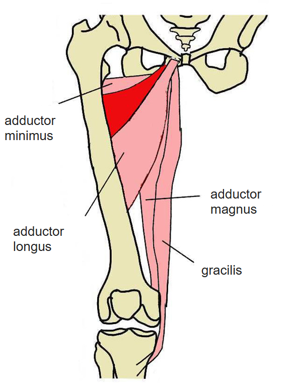
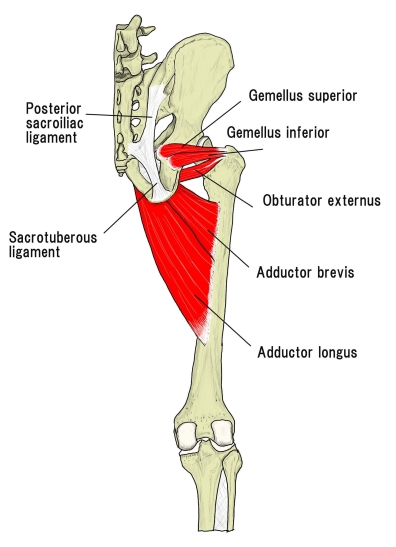
- A roughly triangular adductor lying just deep to the pectineus and adductor longus.
- One of the muscles seen in the femoral triangle; some illustrations show about half of its belly between the pectineus and the adductor longus.
- Pierced near its insertion by the middle perforating artery.
Position in the classification
Lower Limb › Thigh › Adductor group
Illustrations
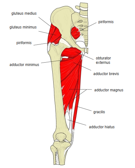
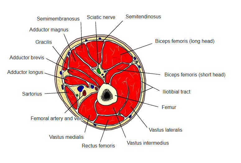
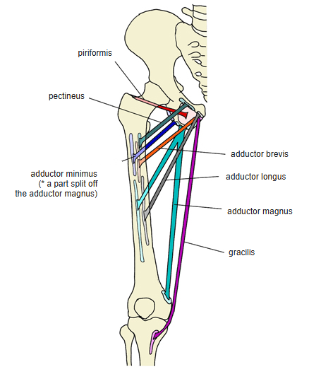
Origin / insertion
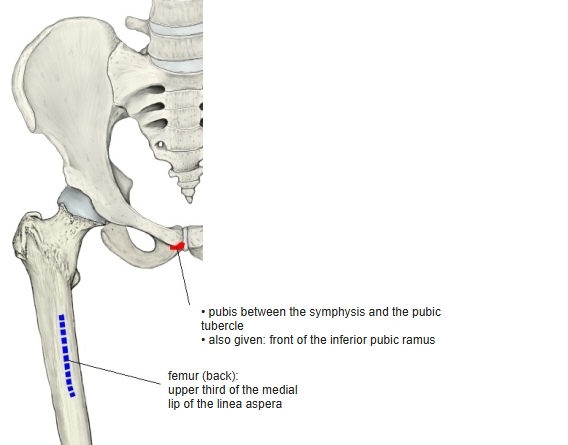
- Origin: pubis
- Between the pubic symphysis and the pubic tubercle
- Also given: anterior surface of the inferior pubic ramus
- Insertion: upper third of the medial lip of the linea aspera of the femur
Action
- Adducts and flexes the thigh; it may rotate the thigh medially or laterally, depending on the position of the hip
- Prometheus: adduction and flexion (up to 70°) at the hip, extension beyond 80° of flexion; steadies the pelvis
Innervation
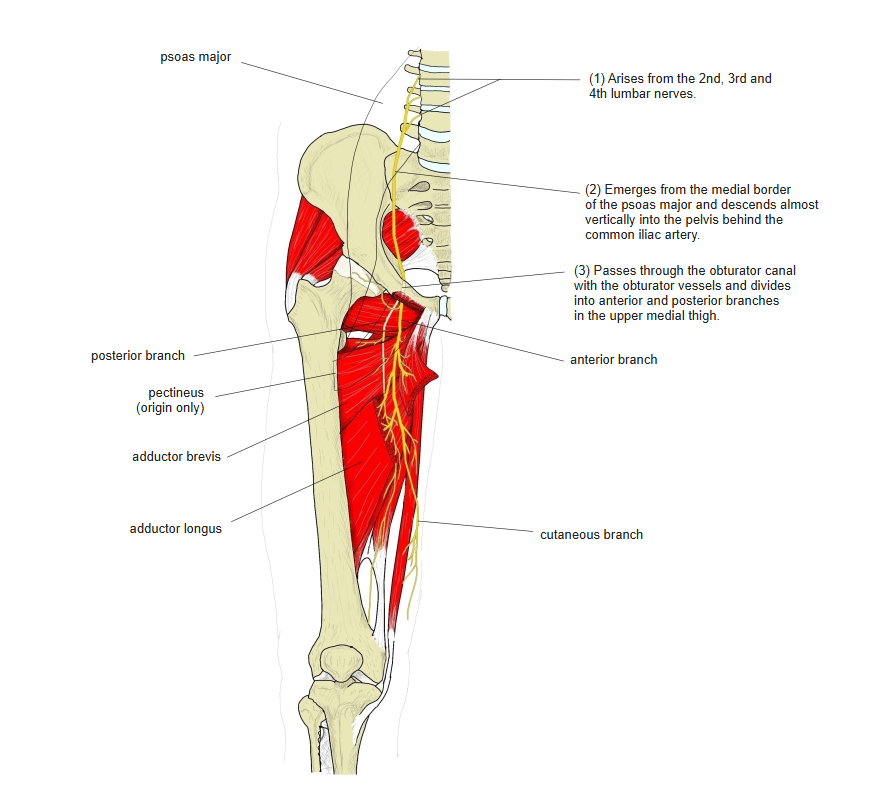
- Obturator nerve (L2–L4); one study found a double supply from both its anterior and posterior branches in about a quarter of cases (Miura et al., 1994)
Blood supply
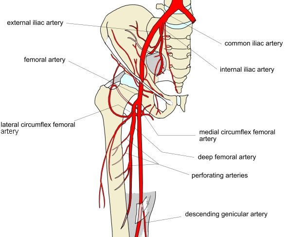
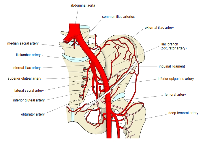
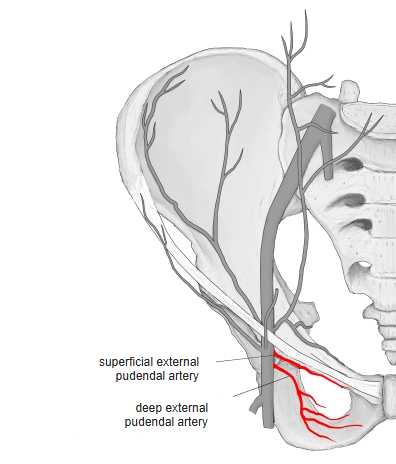
- Deep femoral artery, external pudendal arteries, obturator artery
Anatomical variations
- Sometimes divided in two by a vessel passing through it; may be doubled (Rauber-Kopsch)
Related terms
- Adductor longus
- Adductor magnus
- Pectineus
- Gracilis
- Linea aspera
- Obturator nerve
References and sources
- Further reading: Adductor brevis muscle, Wikipedia.
- Miura et al. The true nature of the adductor brevis dually innervated by the anterior and posterior branches of the obturator nerve in humans. Okajimas Folia Anatomica Japonica 71(2–3), 1994.