Latin: auris media
Overview
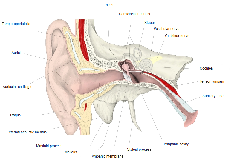
- The air-filled part of the ear between the tympanic membrane and the internal ear, inside the temporal bone.
- Consists of the tympanic cavity with the three auditory ossicles, the auditory tube and the mastoid antrum and mastoid cells.
- Transmits the vibrations of the tympanic membrane to the oval window and increases their pressure.
- Glossary entry: middle ear.
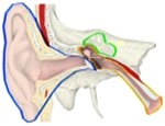
External, middle and internal ear 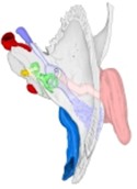
Parts of the ear (superior view)
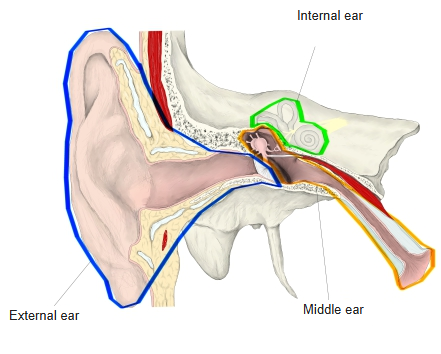
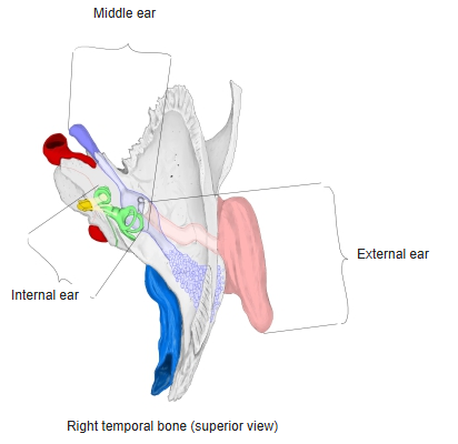
Parts
| Part | Description |
|---|---|
| Tympanic cavity | Narrow air space behind the tympanic membrane; contains the ossicles, two muscles and the chorda tympani |
| Auditory ossicles | Malleus, incus and stapes, linked by synovial joints |
| Auditory tube | Tube from the front of the tympanic cavity to the nasopharynx; equalises air pressure |
| Mastoid antrum and mastoid cells | Air spaces in the mastoid process behind the tympanic cavity, reached through the aditus |
- Muscles: tensor tympani (to the malleus, mandibular nerve) and stapedius (to the stapes, facial nerve).
- The whole middle ear is lined by mucous membrane continuous with that of the nasopharynx – so throat infections can spread to the middle ear (otitis media).
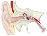
Ear, coronal section
References and sources
- Further reading: Middle ear, Wikipedia.