Latin: rami musculares nervorum intercostalium

Overview

  • Motor branches of the intercostal nerves (and the subcostal nerve) to the muscles of the chest wall, the front and side of the abdominal wall and some posterior thoracic muscles.
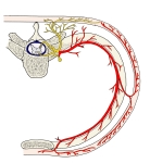
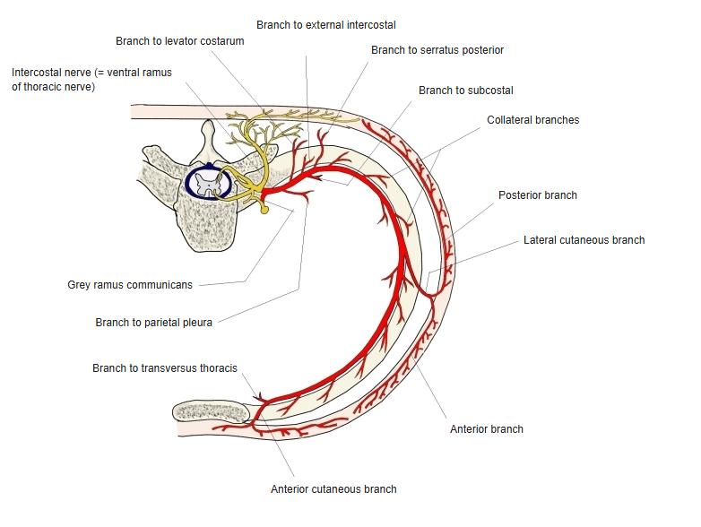
  • Cross-section of the thoracic wall showing an intercostal nerve with its muscular, collateral, lateral cutaneous, anterior cutaneous and pleural branches and the grey ramus communicans
    Branches of an intercostal nerve

Muscles supplied

RegionMuscles
Chest wallInternal intercostals, external intercostals, subcostales, transversus thoracis
Abdominal wallInternal oblique, external oblique, rectus abdominis, transversus abdominis, pyramidalis
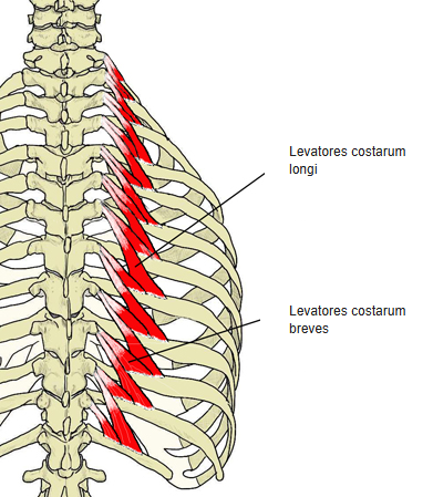
OthersSerratus posterior superior, serratus posterior inferior, levatores costarum
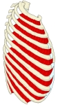
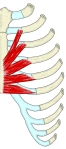
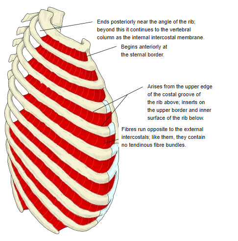
  • Lateral view of the rib cage with the internal intercostal muscles in red, with notes on their origin, insertion and fibre direction
    Internal intercostal muscles
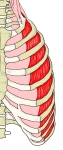
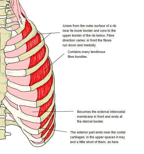
  • Anterolateral view of the rib cage with the external intercostal muscles in red, with notes on their origin, fibre direction and the external intercostal membrane
    External intercostal muscles
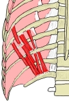
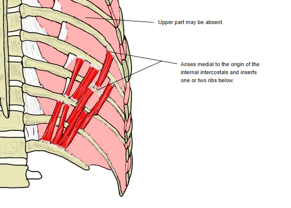
  • Inner surface of the posterior chest wall with the subcostal muscles in red, crossing one or two ribs
    Subcostal muscles
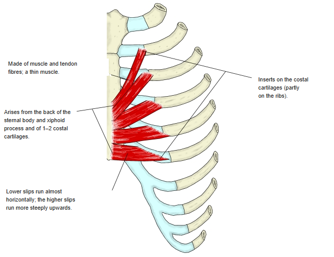
  • Inner surface of the anterior chest wall with transversus thoracis in red, running from the sternum to the costal cartilages
    Transversus thoracis
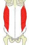
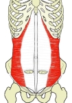
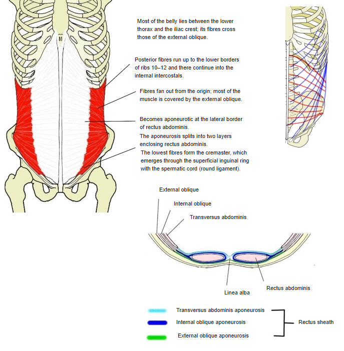
  • Front view of the abdomen with the internal oblique in red, a cross-section of the abdominal wall showing the layers of the rectus sheath, and a side view of the fibre directions
    Internal oblique and rectus sheath
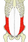
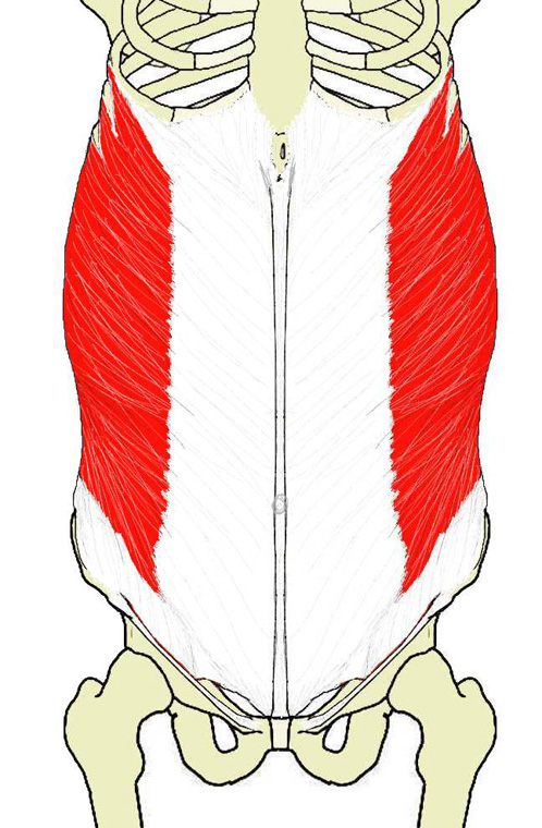
  • Front view of the abdomen with transversus abdominis in red
    Transversus abdominis
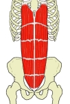
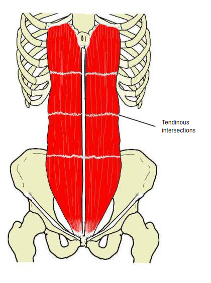
  • Front view of the abdomen with rectus abdominis in red and its tendinous intersections
    Rectus abdominis
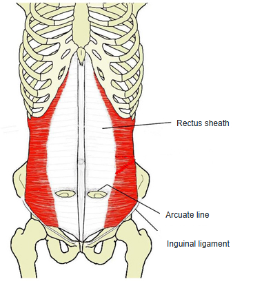
  • Front view of the abdominal wall showing the rectus sheath, the arcuate line and the inguinal ligament
    Rectus sheath
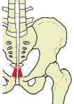
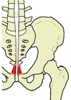
  • Front view of the lower abdomen and pelvis with the small pyramidalis muscles in red above the pubic symphysis
    Pyramidalis
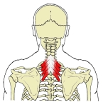
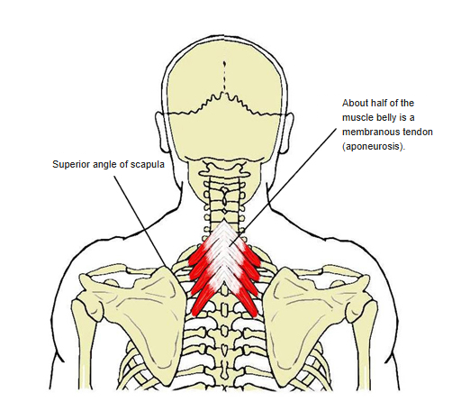
  • Back view of the upper thorax with serratus posterior superior in red, between the cervicothoracic spinous processes and the upper ribs
    Serratus posterior superior
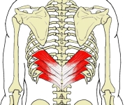
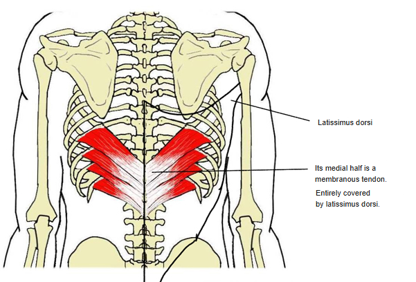
  • Back view of the lower thorax with serratus posterior inferior in red, deep to latissimus dorsi
    Serratus posterior inferior
  • Back view of the thoracic spine and ribs with the long and short levatores costarum muscles in red
    Levatores costarum

References and sources

  1. Further reading: Muscular branches of intercostal nerves, Wikipedia.