Latin: Arteria maxillaris
Overview
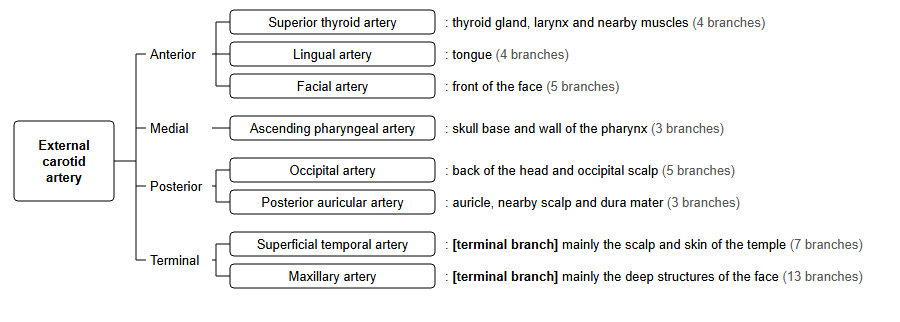
- The larger of the two terminal branches of the external carotid artery; the other is the superficial temporal artery.
- Supplies the deep structures of the face: jaws and teeth, muscles of mastication, nasal cavity, palate and the dura mater.
- Runs forward from behind the neck of the mandible, through the infratemporal fossa, to the pterygopalatine fossa.
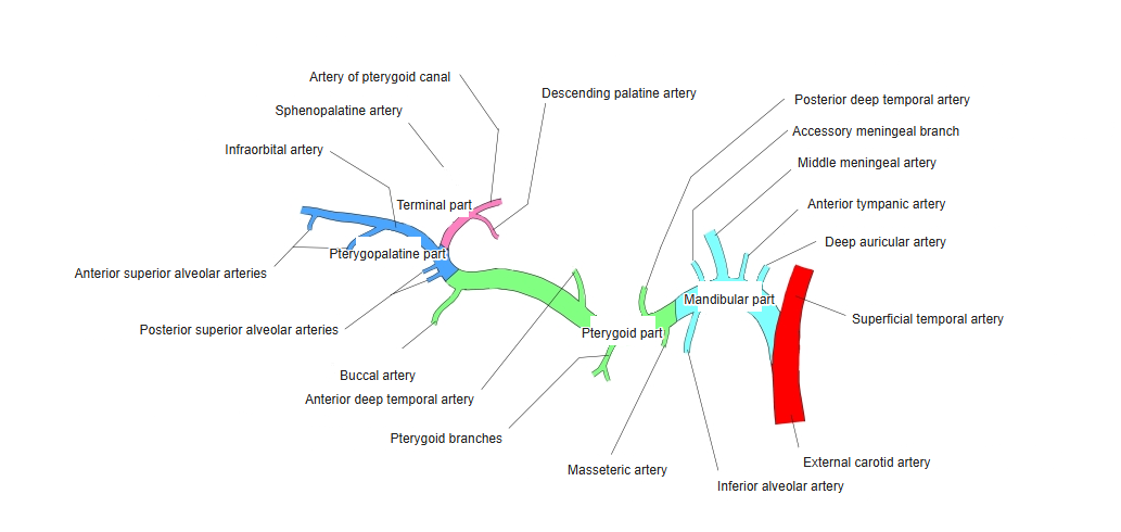
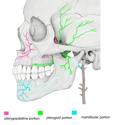
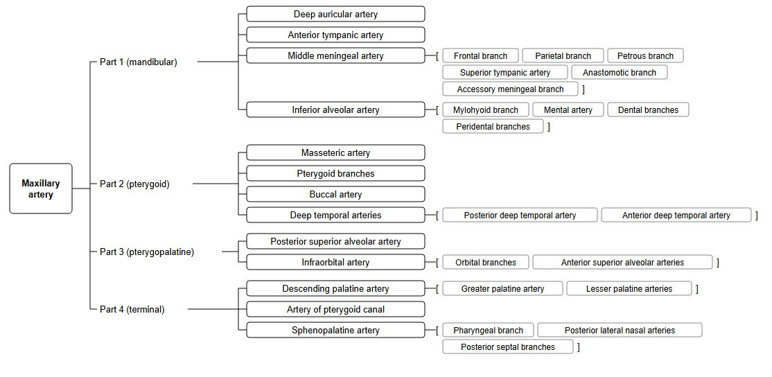
- Usually described in three parts (mandibular, pterygoid, pterygopalatine). Some sources count the terminal branches in the pterygopalatine fossa as a fourth part.
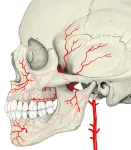
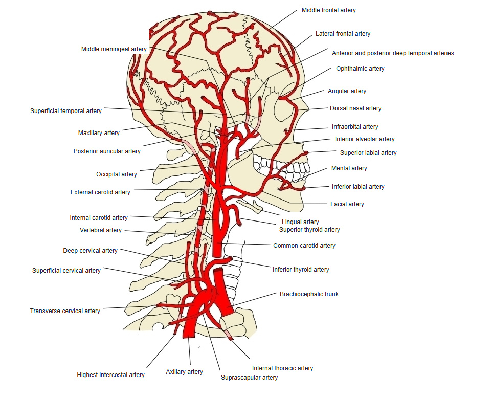
Maxillary artery and its branches (lateral view) 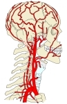
Arteries of the head and neck 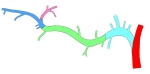
Branches by part (diagram)
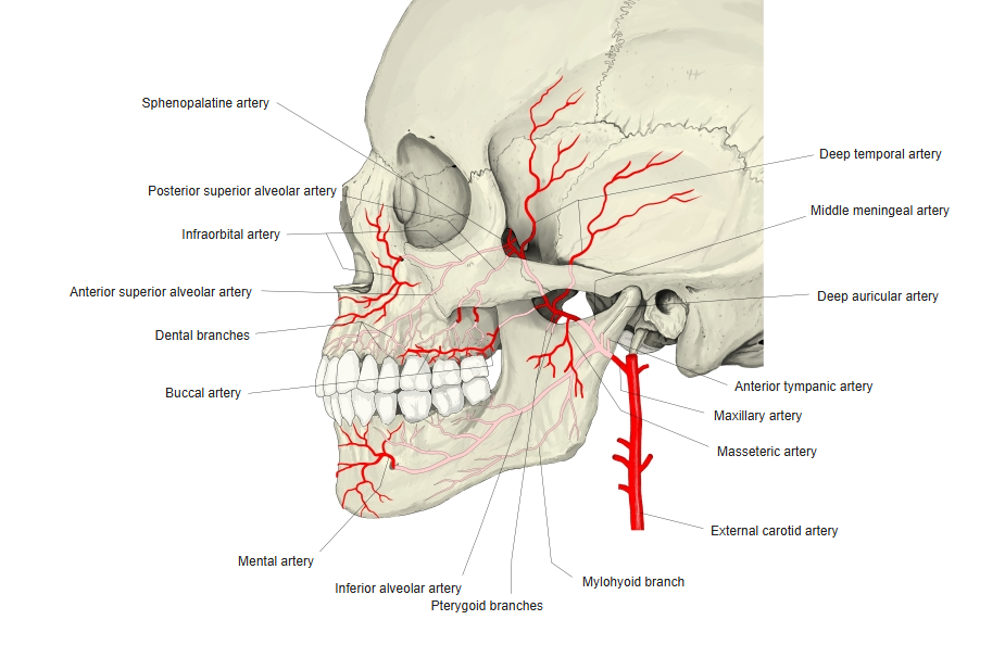

Origin
- Arises from the external carotid artery behind the neck of the mandible, embedded at first in the parotid gland.
- The external carotid artery ends here by dividing into the maxillary and superficial temporal arteries.
Course
- Mandibular part: passes forward between the neck of the mandible and the sphenomandibular ligament, along the lower border of the lateral pterygoid.
- Pterygoid part: ascends obliquely forward, either superficial or deep to the lateral pterygoid muscle.
- Pterygopalatine part: enters the pterygopalatine fossa through the pterygomaxillary fissure and divides into its terminal branches.
Branches
| Part | Branch | Main area supplied |
|---|---|---|
| Mandibular | Deep auricular artery | External acoustic meatus, temporomandibular joint |
| Mandibular | Anterior tympanic artery | Tympanic membrane, tympanic cavity |
| Mandibular | Middle meningeal artery | Dura mater and bones of the calvaria |
| Mandibular | Accessory meningeal branch | Dura mater, trigeminal ganglion, nearby muscles |
| Mandibular | Inferior alveolar artery (gives the mylohyoid branch and the mental artery) | Lower teeth, mandible, chin |
| Pterygoid | Deep temporal arteries (anterior and posterior) | Temporalis |
| Pterygoid | Masseteric artery | Masseter |
| Pterygoid | Pterygoid branches | Medial and lateral pterygoid muscles |
| Pterygoid | Buccal artery | Buccinator, cheek |
| Pterygopalatine | Posterior superior alveolar artery | Upper molars and premolars, maxillary sinus, gums |
| Pterygopalatine | Infraorbital artery (gives the anterior superior alveolar arteries) | Lower eyelid, upper lip, upper front teeth |
| Pterygopalatine | Descending palatine artery (greater and lesser palatine arteries) | Hard and soft palate, palatine tonsil |
| Pterygopalatine | Artery of the pterygoid canal | Upper pharynx, auditory tube |
| Pterygopalatine | Sphenopalatine artery | Nasal cavity (lateral wall and septum) |
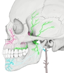
Three parts of the maxillary artery
Clinical notes
- Tearing of the middle meningeal artery (for example in a fracture of the temporal region) can cause an epidural hematoma.
- The sphenopalatine artery is the main source of posterior nosebleeds (epistaxis).
- The inferior alveolar artery runs with the inferior alveolar nerve; this is relevant to the inferior alveolar nerve block in dentistry.
References and sources
- Further reading: Maxillary artery, Wikipedia.