Pterygopalatine fossa (Latin: fossa pterygopalatina)
Also called the sphenopalatine fossa
- Overview
- Bones forming it
- Communications
- Reference websites
- (Wikipedia, websites with illustrations)
- Where it is found
- Related terms
Overview
The pterygopalatine fossa is a cone-shaped space that lies medial to the infratemporal fossa, just below the apex of the orbit, and through which many nerves and vessels pass.
Infratemporal fossa: the space that continues downwards and medially from the temporal fossaCone-shaped: it narrows downwards
It is an important crossroads that connects other spaces of the skull: the middle cranial fossa, the orbit and the nasal cavity, as well as the oral cavity, the nasopharynx and the infratemporal fossa. It contains the pterygopalatine ganglion, the maxillary nerve and the last part of the maxillary artery with its veins; local anaesthetic injected into it can block the whole maxillary nerve.
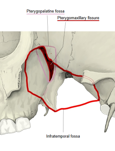
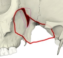 Infratemporal and pterygopalatine fossae
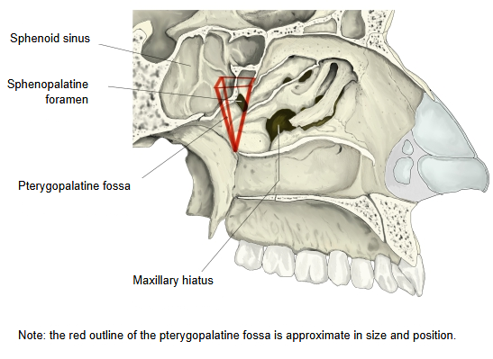
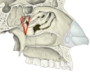 Pterygopalatine fossa seen from the nasal cavity
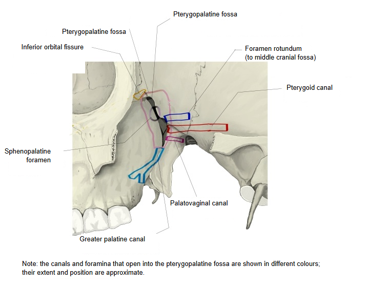
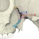 Openings of the pterygopalatine fossa
Bones forming it
The pterygopalatine fossa is bounded by the following bones.
| Front | · The back border (infratemporal surface) of the body of the maxilla (maxilla), near the maxillary tuberosity · Part of the orbital process of the palatine bone |
| Back | · The pterygoid process (root and lateral plate) and the adjoining front of the greater wing of the sphenoid bone |
| Medial | · The perpendicular plate of the palatine bone, with its orbital and sphenoidal processes |
| Lateral | · No wall: a triangular gap, the pterygomaxillary fissure, opening into the infratemporal fossa; in life its lower half is closed over by periosteum. |
| Above | · The undersurface of the body of the sphenoid (in front and above, the fossa opens into the inferior orbital fissure and has no wall of its own) |
| Below | · The body of the maxilla · The pterygoid process of the sphenoid · The pyramidal process of the palatine bone (here the fossa narrows into the upper opening of the greater palatine canal) |
Communications
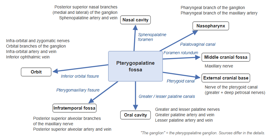
The pterygopalatine fossa communicates in every direction - front and back, medially and laterally, above and below - with other spaces of the skull, and various nerves and vessels pass through it. The nerves and vessels in the table follow a standard atlas of head and neck anatomy; sources differ in some details.
| Direction | Route | Leads from / to | Nerves and vessels |
|---|---|---|---|
| Front, above | Inferior orbital fissure | Orbit | Nerves: infra-orbital nerve, zygomatic nerve, orbital branches (from the pterygopalatine ganglion, on the maxillary nerve) Vessels: infra-orbital artery and vein, connections with the inferior ophthalmic vein |
| Back | Foramen rotundum | Middle cranial fossa | · Maxillary nerve |
| Pterygoid canal | External cranial base (region of the foramen lacerum) | · Nerve of the pterygoid canal (the greater and deep petrosal nerves combined) · Artery and vein of the pterygoid canal | |
| Back, medially | Palatovaginal canal | Nasopharynx | · Pharyngeal branch of the pterygopalatine ganglion · Pharyngeal branch of the maxillary artery |
| Medially | Sphenopalatine foramen | Nasal cavity | · Medial and lateral posterior superior nasal branches of the pterygopalatine ganglion · Sphenopalatine artery and vein Sources differ: a Japanese anatomy textbook names the posterior superior nasal nerves; a standard atlas the lateral and medial posterior superior nasal branches of the maxillary nerve and the sphenopalatine artery (with its vein); English Wikipedia the posterior superior lateral nasal nerves, the nasopalatine nerve and the sphenopalatine artery and vein. |
| Laterally | Pterygomaxillary fissure | Infratemporal fossa | · The maxillary artery entering the fossa · Posterior superior alveolar branches of the maxillary nerve and the posterior superior alveolar artery and vein leaving it (this row may be less certain) |
| Below | Greater palatine canal | Oral cavity | · Greater palatine nerve, lesser palatine nerve · Greater palatine artery and vein |
| Lesser palatine canals (branching from the greater palatine canal) | · Lesser palatine nerve · Lesser palatine artery (a terminal branch of the descending palatine artery) and vein |
Reference websites
[Wikipedia]
· Pterygopalatine fossa, Wikipedia.
· Pterygopalatine ganglion, Wikipedia.
[Websites with illustrations]
· Boundaries of the pterygopalatine fossa (AnatomyQA)

· Openings of the pterygopalatine fossa (AnatomyQA)

· Pterygopalatine fossa (NotesMed)

Where it is found
Related terms
- Infratemporal fossa
- Pterygopalatine ganglion
- Foramen rotundum
- Sphenopalatine foramen
- Greater palatine canal
- Pterygoid process
- Maxillary nerve
References and sources
- Further reading: Pterygopalatine fossa, Wikipedia.
- Further reading: Pterygopalatine ganglion, Wikipedia.
- Federative International Programme for Anatomical Terminology (FIPAT). Terminologia Anatomica, 2nd ed. (TA2), 2019.