Infratemporal fossa (Latin: fossa infratemporalis)
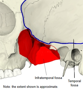
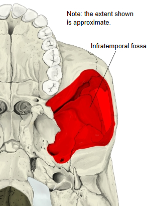
The infratemporal fossa is the space that continues downwards and medially from the temporal fossa. It lies on the side of the skull base, below the zygomatic arch and deep to the ramus of the mandible, behind the maxilla.
· It holds the pterygoid muscles and the main branches of the mandibular nerve and the maxillary artery, which pass on to the jaws, teeth, tongue and face.
More details
Walls
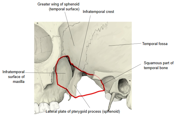
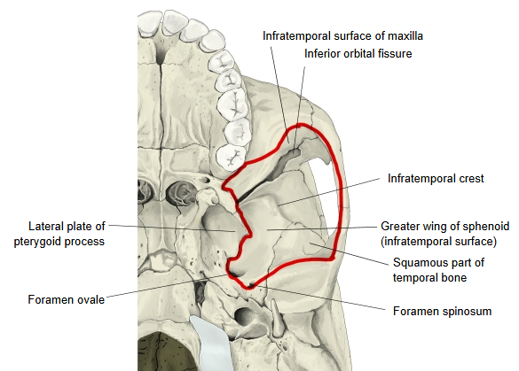
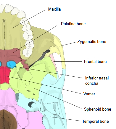
Three walls of the infratemporal fossa can be recognised on the skull:
- Roof : sphenoid bone (infratemporal surface of the greater wing) and temporal bone (a small part of the squamous part); pierced by the foramen ovale and the foramen spinosum
- Anterior wall : maxilla (infratemporal surface)
- Medial wall : sphenoid bone (lateral plate of the pterygoid process)
- Lateral, posterior, inferior : no bony wall: laterally lies the ramus of the mandible; behind lie the styloid process and the carotid sheath; below, the fossa ends where medial pterygoid attaches to the mandible
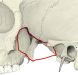 Bony walls (lateral view)
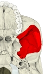 Infratemporal fossa (inferior view)
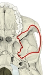 Bony walls (inferior view)
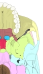 Bones around the fossa (inferior view)
Contents
The infratemporal fossa contains the following:
Muscles: medial pterygoid and lateral pterygoid. Some sources also include the lower part of temporalis.
Nerves: the mandibular nerve and its branches (including the inferior alveolar, lingual, buccal and auriculotemporal nerves), the chorda tympani joining the lingual nerve, and the otic ganglion.
Vessels: the maxillary artery and its branches, and the pterygoid plexus of veins.
Some sources also list the coronoid process of the mandible, to which temporalis attaches.
Communications
In front of, medial to and above the infratemporal fossa there are foramina and fissures through which it communicates with other spaces of the skull:
- In front : inferior orbital fissure → orbit
- Medially : pterygomaxillary fissure → pterygopalatine fossa
- Above : foramen ovale, foramen spinosum → middle cranial fossa
- Above and laterally : the gap deep to the zygomatic arch → temporal fossa; the infratemporal crest of the greater wing marks the border between the two fossae
Photographs and illustrations
Reference websites
[Wikipedia]
· Infratemporal fossa, Wikipedia.
[Websites with illustrations]
· Skull base from below, with the infratemporal fossa (photograph) (Wikimedia Commons)

· Infratemporal fossa, deep dissection (contents) (Instant Anatomy)

Where it is found
Related terms
- Pterygopalatine fossa
- Greater wing
- Pterygoid process
- Foramen ovale
- Ramus of mandible
- Zygomatic arch
- Maxillary artery
References and sources
- Further reading: Infratemporal fossa, Wikipedia.
- Further reading: Temporal fossa, Wikipedia.
- Federative International Programme for Anatomical Terminology (FIPAT). Terminologia Anatomica, 2nd ed. (TA2), 2019.