Latin: Arteria carotis communis
Overview
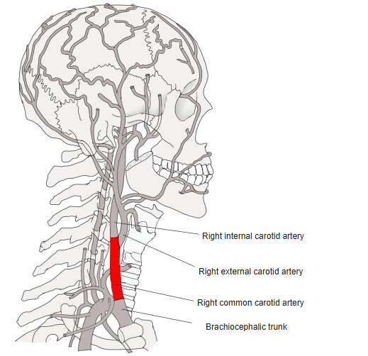
- The main artery of the head and neck, one on each side.
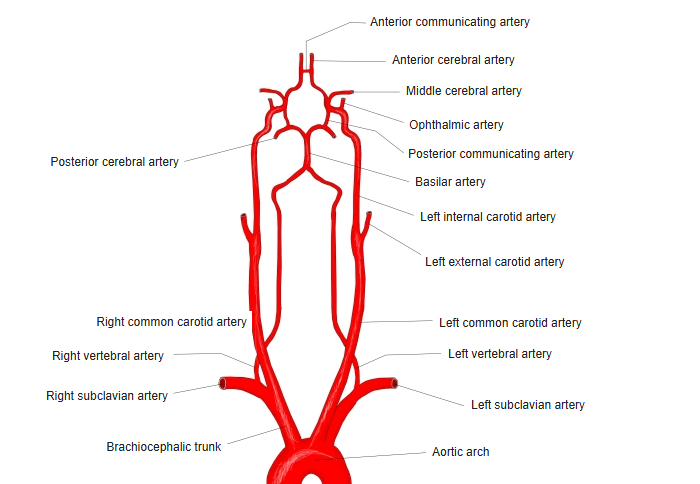
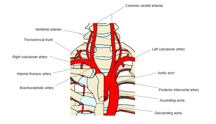
- The right arises from the brachiocephalic trunk; the left arises directly from the aortic arch.
- Divides at about the level of the upper border of the thyroid cartilage (C3–C4) into the internal and external carotid arteries.
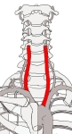
Right and left common carotid arteries 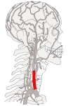
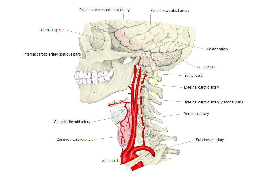
Common carotid artery (side view) 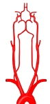
Carotid and vertebral arteries (front view)
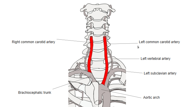
Origin
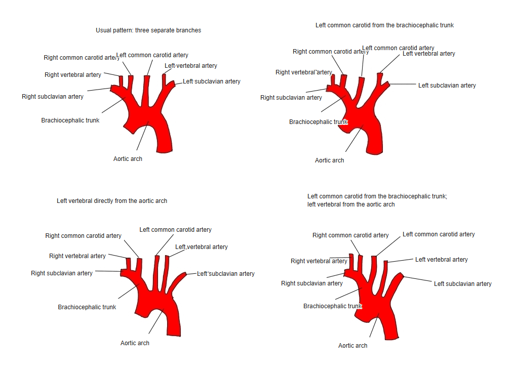
- The origins of the arteries from the aortic arch vary; the most common pattern has three separate branches.
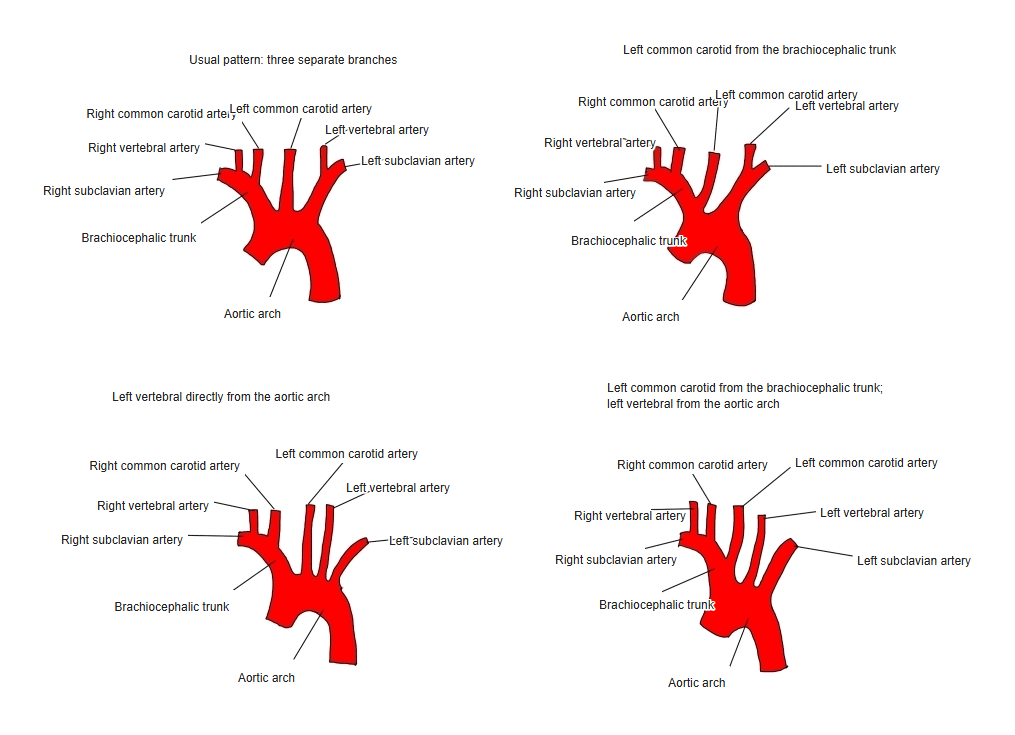
Variations in the branching of the aortic arch 
Branches of the aortic arch 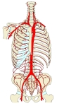
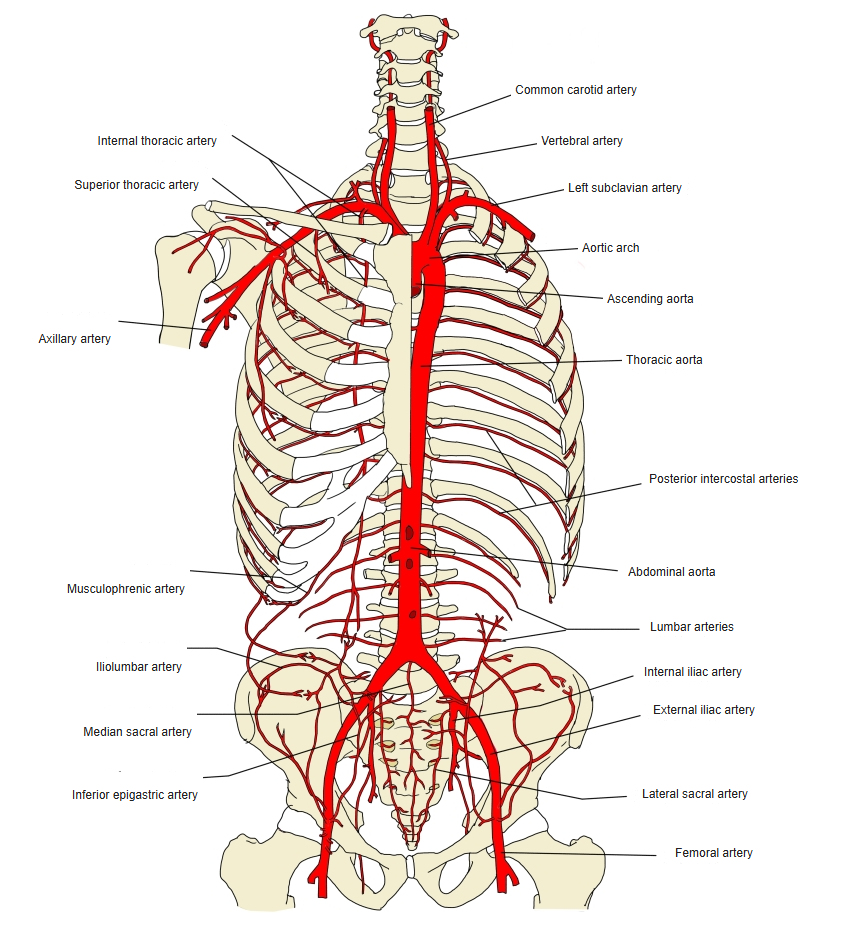
Aorta and its branches in the trunk (front view)
Course
- Ascends in the neck inside the carotid sheath with the internal jugular vein (lateral) and the vagus nerve (behind).
- Has no branches before its division. At the division it widens into the carotid sinus (pressure receptor); the carotid body (chemoreceptor) lies behind it.
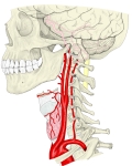
Common, external and internal carotid arteries 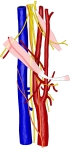
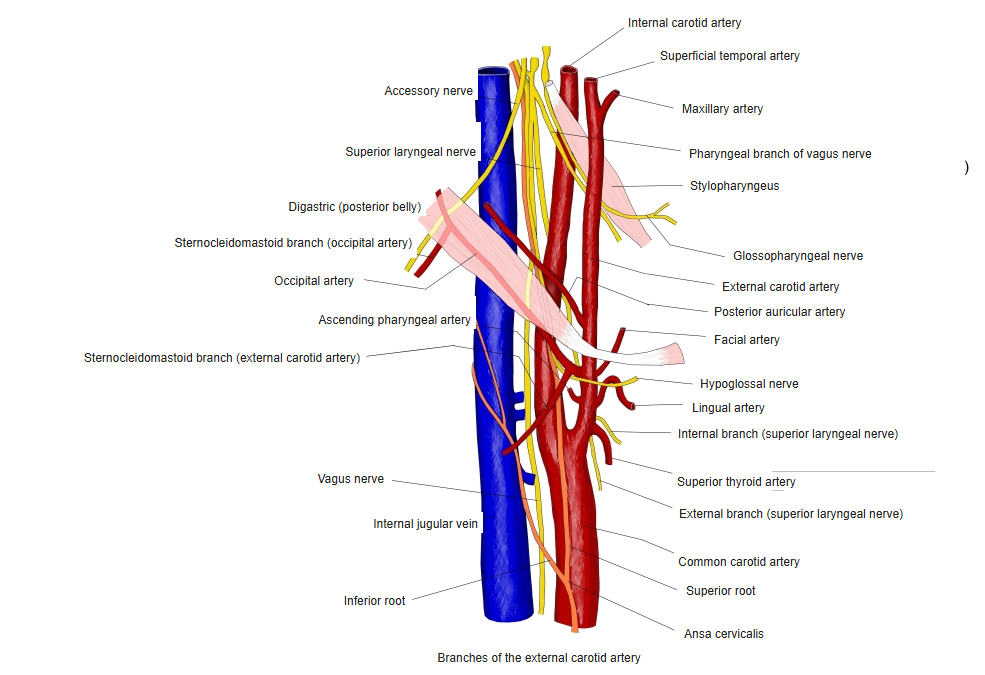
Carotid arteries and nearby nerves (front view) 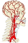
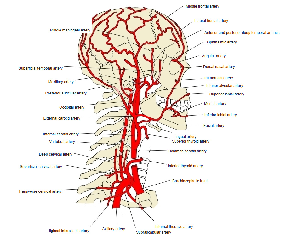
Arteries of the head and neck 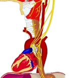
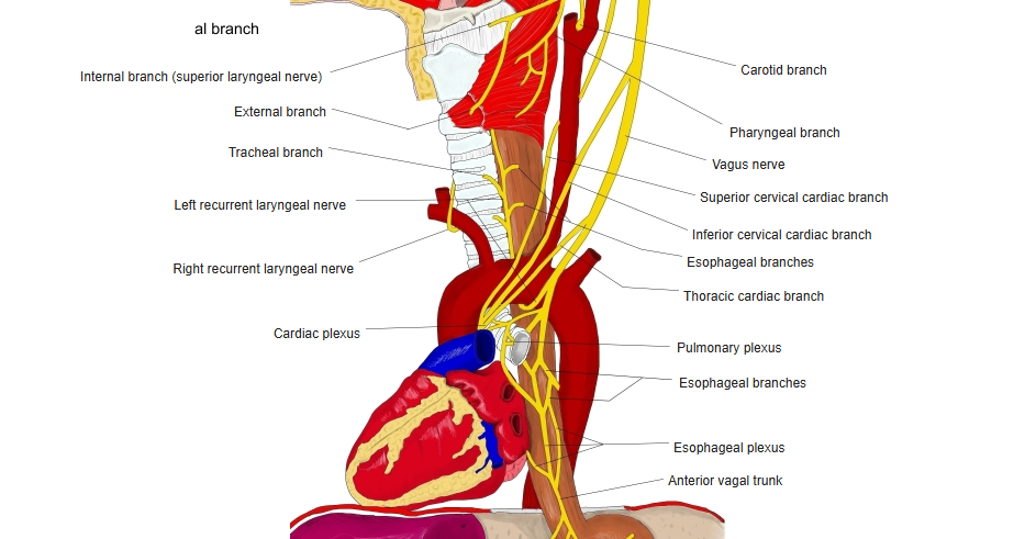
Vagus nerve and the great arteries
Clinical notes
- The carotid pulse can be felt at the front edge of the sternocleidomastoid at the level of the thyroid cartilage.
- Narrowing at the bifurcation (carotid stenosis) is a common cause of stroke.
References and sources
- Further reading: Common carotid artery, Wikipedia.