Latin: Aorta thoracica
Overview
- The part of the descending aorta in the thorax, from the lower border of the 4th thoracic vertebra to the aortic hiatus of the diaphragm (about T12).
- Lies in the posterior mediastinum, first on the left side of the vertebral bodies and then moving toward the midline.
- Continues below the diaphragm as the abdominal aorta.
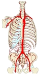
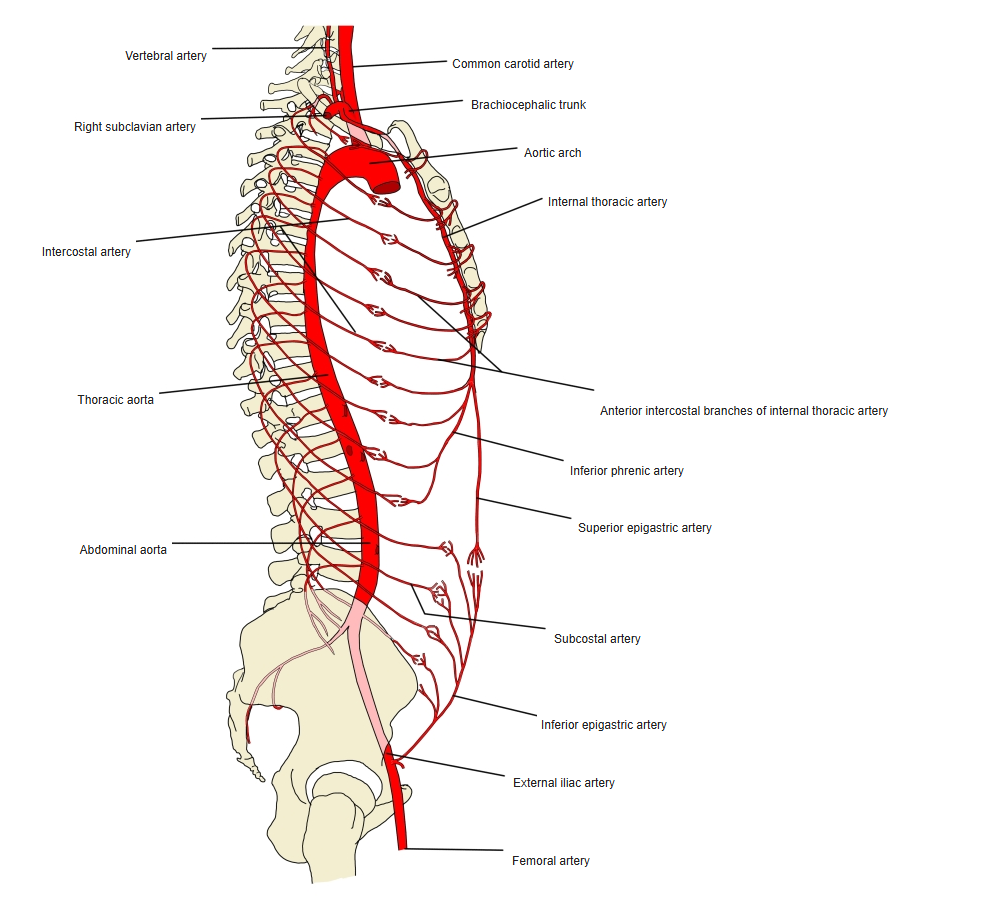
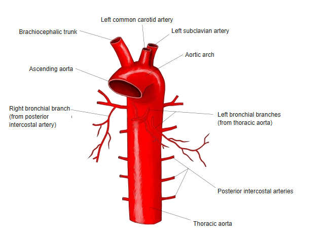
Aorta and its branches in the trunk (front view) 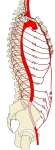
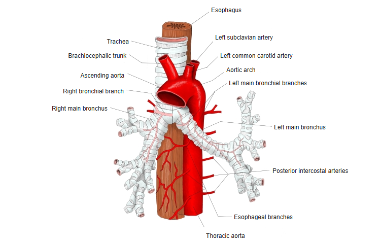
Aorta and its branches in the trunk (side view) 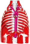
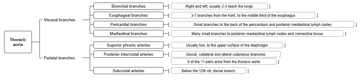
Thoracic aorta and its branches
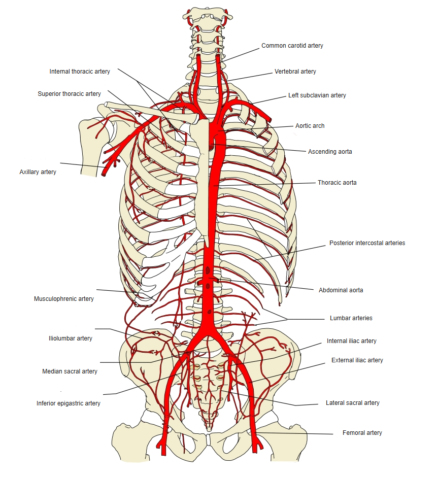
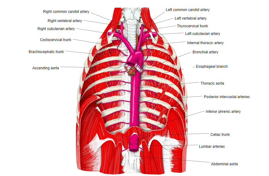
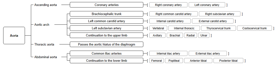
Branches
| Group | Branches | Supply |
|---|---|---|
| Visceral branches | Bronchial branches | Bronchi and lung tissue (usually two left bronchial arteries; the right usually arises from the 3rd posterior intercostal artery) |
| Esophageal branches | Thoracic part of the esophagus | |
| Mediastinal and pericardial branches | Lymph nodes and connective tissue of the posterior mediastinum; back of the pericardium | |
| Parietal branches | Posterior intercostal arteries (3rd–11th) | Intercostal spaces, back muscles, spinal cord |
| Subcostal arteries | Below the 12th ribs | |
| Superior phrenic arteries | Upper surface of the diaphragm |
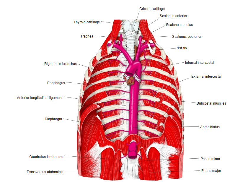
Thoracic aorta and nearby structures 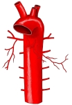
Bronchial branches 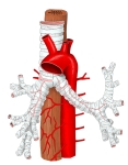
Esophageal branches 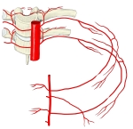
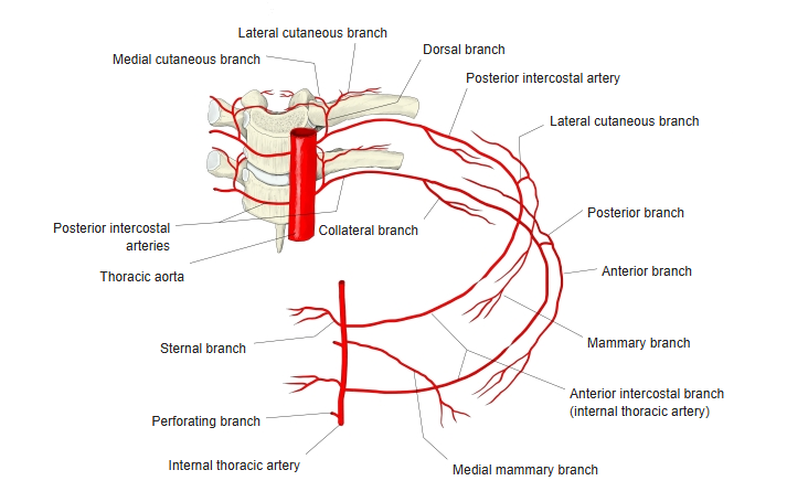
Intercostal arteries in one intercostal space
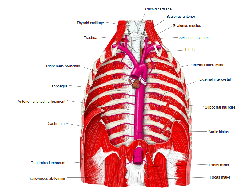
Relations
- The esophagus lies to its right above and passes in front of it lower down.
- The azygos vein and the thoracic duct lie on its right side.
References and sources
- Further reading: Thoracic aorta, Wikipedia.