Latin: Vena jugularis interna
Overview
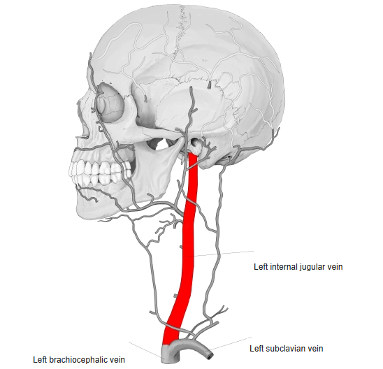
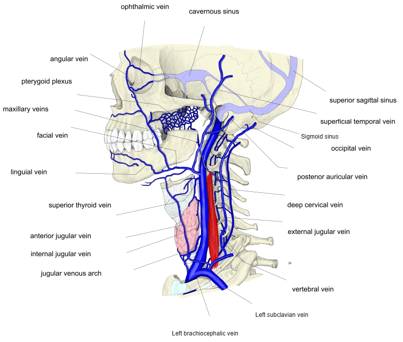
- The main vein of the neck. It collects blood from the brain, the inside of the skull, and the deep parts of the face and neck.
- Begins at the jugular foramen at the base of the skull as the continuation of the sigmoid sinus; its first part is dilated as the superior bulb.
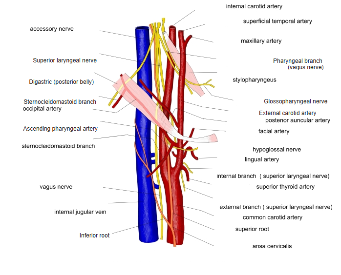
- Runs down in the carotid sheath, lateral to the internal carotid and then the common carotid artery, with the vagus nerve between and behind them.
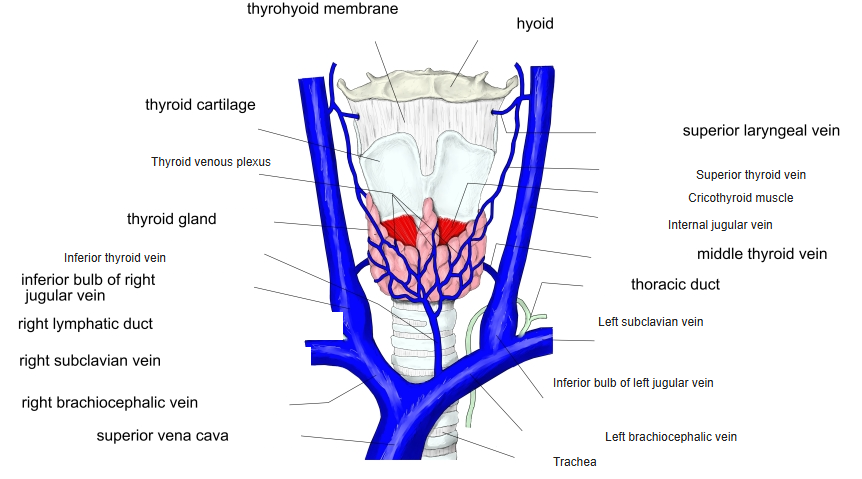
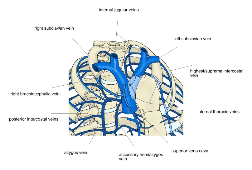
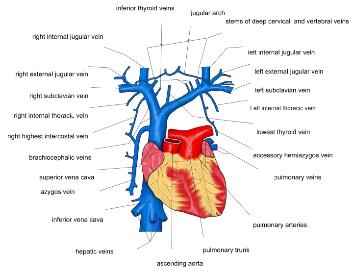
- Ends behind the sternal end of the clavicle by joining the subclavian vein to form the brachiocephalic vein; just above this it has a second dilatation, the inferior bulb, with a valve.
Internal jugular vein 
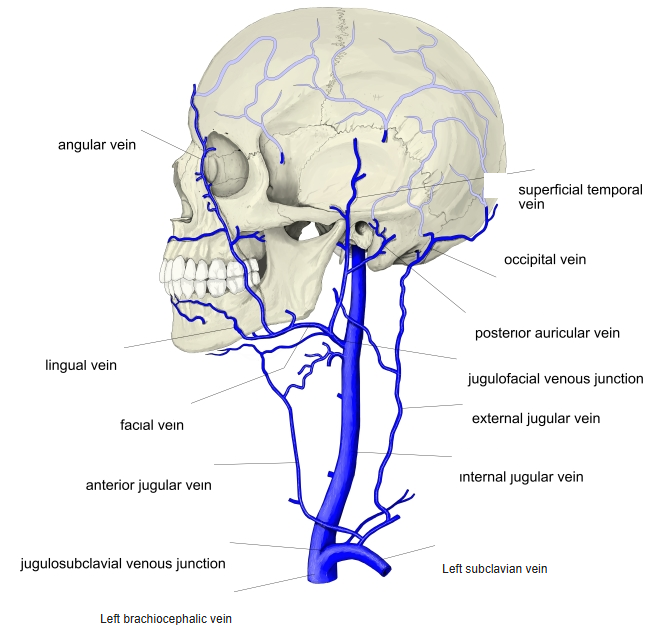
Veins of the head and neck (side view)
Tributaries
| Region | Tributaries |
|---|---|
| Inside the skull and at the jugular foramen | Inferior petrosal sinus, vein of the cochlear canaliculus |
| Face and tongue | Facial vein / common facial vein, lingual vein |
| Pharynx | Pharyngeal veins |
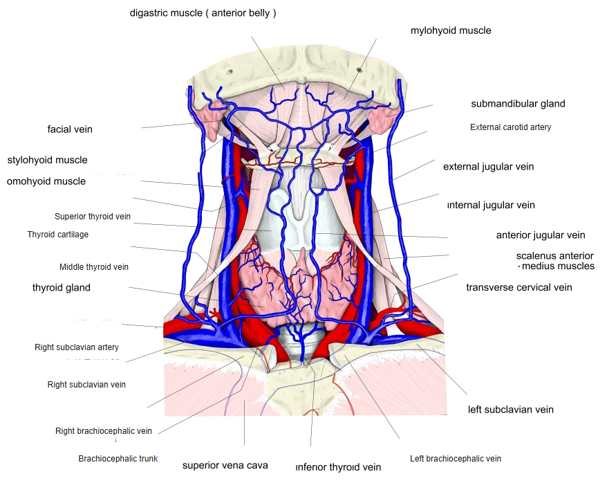
| Larynx and thyroid | Superior and middle thyroid veins, superior laryngeal vein (via the superior thyroid vein) |
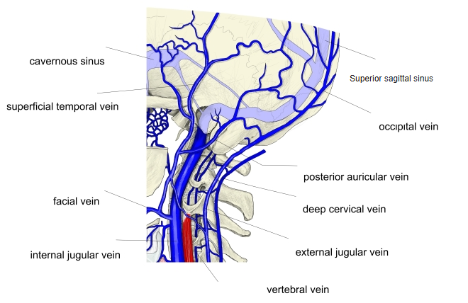
| Muscles | Sternocleidomastoid vein; sometimes the occipital vein |
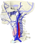
Tributaries of the jugular veins 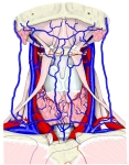
Veins of the front of the neck 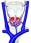
Veins of the thyroid gland and larynx 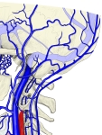
Veins of the back of the head and neck
Relations
- Covered by the sternocleidomastoid for most of its course.
- Above: close to the cranial nerves IX–XII at the skull base.
- Below: the subclavian artery and (on the left) the thoracic duct lie near its end.
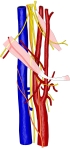
Internal jugular vein and nearby structures 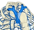
Formation of the superior vena cava 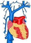
Superior vena cava and its tributaries 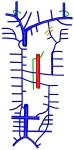
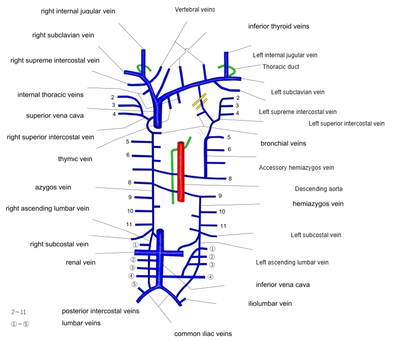
Veins of the thorax (diagram)
Clinical notes
- Commonly used for central venous catheters.
- The jugular venous pulse in the neck reflects pressure in the right atrium.
- Deep cervical lymph nodes lie along it, so it is a landmark in neck dissection.
References and sources
- Further reading: Internal jugular vein, Wikipedia.