Tracheal cartilages (Latin: cartilagines tracheales)
Also called Tracheal rings
· They are normally elastic, but with age the tracheal cartilages tend to calcify and become stiffer.
· With age two (or more) of the cartilages may fuse together.
the trachea
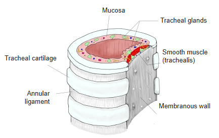
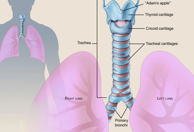
- Tracheal cartilages : 16–20 C-shaped rings of hyaline cartilage, each about 3–4 mm high.
- Annular ligaments : the parts of the connective-tissue (fibroelastic) membrane around the cartilages that stretch between them.
- Mucosa : well developed and lining the lumen, without folds. It contains the tracheal glands, vessels and nerves.
- Membranous wall : the back wall, facing the esophagus. It is made mainly of smooth muscle (the trachealis) and contains large tracheal glands.
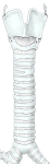 Trachea (anterior view)
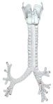 Trachea and bronchi (posterior view)
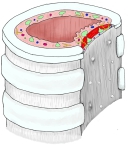 Trachea (cross-section)
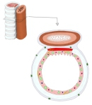 Trachea and esophagus (cross-section)
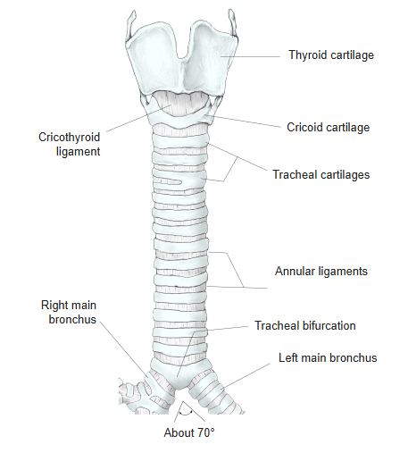
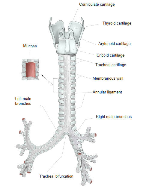
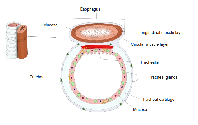
The cartilages support the trachea and keep it open, yet still let it move, stretch and bend with breathing and with movements of the neck. The C-shaped rings lie one on top of another, open where the trachea is nearest the esophagus. The first cartilage is broad and is joined to the cricoid cartilage by the cricotracheal ligament; it may be split at one end or fused with the next. The last cartilage is thick in the middle, and its triangular lower edge bends down and back between the two main bronchi to form the carina at the tracheal bifurcation. Sources differ on the number of rings (from 15 to about 22) and on their exact size.
Reference websites
[Wikipedia]
· Trachea, Wikipedia.
[Websites with illustrations]
· Trachea: anatomy and function (Kenhub)
· Cartilages of the larynx, trachea and bronchi, front view (Gray's Anatomy) (Wikimedia Commons)

· Trachea (figure) (WebMD)
Where it is found
Related terms
References and sources
- Further reading: Trachea, Wikipedia.
- Federative International Programme for Anatomical Terminology (FIPAT). Terminologia Anatomica, 2nd ed. (TA2), 2019.