Alveolar pores (Latin: pori alveolares)
Also called the pores of Kohn or the interalveolar pores or the singular alveolar pore
· Also called the pores of Kohn, after the German pathologist Hans Nathan Kohn, who described them in 1893.
Reference websites
[Wikipedia]
· Pores of Kohn, Wikipedia.
· Pulmonary alveolus, Wikipedia.
[Websites with illustrations]
· Alveoli (illustration) (GetBodySmart)
· Alveoli and pores (image) (Radiopaedia)
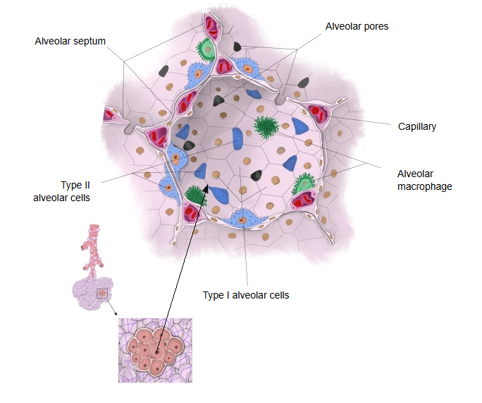
· The edge of each pore is usually formed partly by type II alveolar cells, the cells that make surfactant.
· They are absent or very few in newborns and develop gradually during childhood (from about 3–4 years of age, according to some sources such as Wikipedia; other studies find them earlier), together with the canals of Lambert, as the alveolar septa become thinner; they become more numerous with age.
· Fluid, bacteria, fibrin and other materials pass between alveoli through the pores, and alveolar macrophages can move through them too. This helps explain how lobar pneumonia spreads through a whole lobe; their poor development in young children is thought to help keep pneumonia localised ("round pneumonia").
· By connecting neighbouring alveoli, the pores equalise the pressure between them. Together with the canals of Lambert they provide collateral ventilation: air can reach alveoli whose own airway is blocked, which helps prevent alveolar collapse.
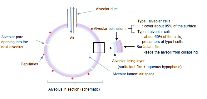
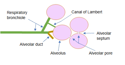
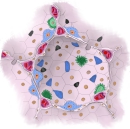 Alveolar wall and pores
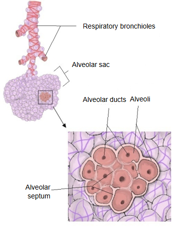
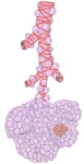 Respiratory bronchioles and alveoli


Where it is found
Related terms
References and sources
- Further reading: Pores of Kohn, Wikipedia.
- Further reading: Pulmonary alveolus, Wikipedia.
- Federative International Programme for Anatomical Terminology (FIPAT). Terminologia Anatomica, 2nd ed. (TA2), 2019.