Gingiva (Latin: gingiva)
Also called the gums
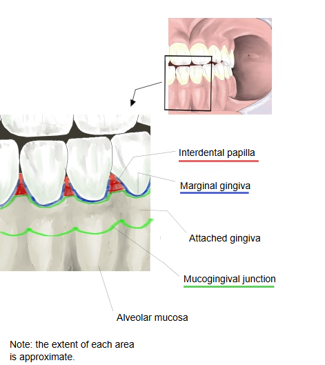
- Attached gingiva : the immovable part that makes up most of the gingiva. Firm and often stippled, it is bound by the gingival fibres to the cementum and to the periosteum of the alveolar bone. Widest in the incisor region, narrowest near the first premolars.
- Interdental gingiva : the gingiva that, seen from the front, fills the space between neighbouring teeth below their contact point as a triangle: the interdental papilla. In the back teeth it has a saddle-shaped dip, the col, covered by non-keratinized epithelium.
- Free gingiva : the unattached collar around the neck of the tooth, about 0.5-2 mm wide, that forms the outer wall of the gingival sulcus. It follows the scalloped line of the cementoenamel junction; also called the marginal gingiva.
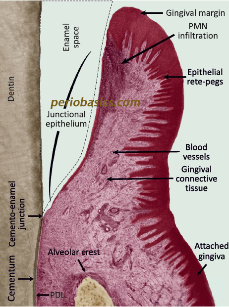
Most of the gingiva is firmly bound to the bone of the alveolar processes, so it resists the friction of chewing; its surface is keratinized or parakeratinized. It forms a seal around each tooth: at the bottom of the shallow gingival sulcus the junctional epithelium attaches to the tooth surface.
Boundaries: a faint free gingival groove may separate the free gingiva from the attached gingiva; it is seen in about half of people. The mucogingival junction separates the attached gingiva from the redder, loose and movable alveolar mucosa. On the palate there is no such junction: the gingiva blends into the mucosa of the hard palate. Sources differ on the exact limits of each part, and on whether the interdental papilla counts as a part of the free gingiva or as a separate part.
Healthy gingiva is coral pink, firm and does not bleed; it may be darker where melanin is present. Redness, swelling and bleeding suggest gingivitis, usually caused by dental plaque.
- Gingiva : the part of the oral mucosa around the teeth, the 'gums'. Includes the attached gingiva, the interdental papillae and the free gingiva.
- Periodontal ligament : the fibrous connective tissue, with vessels and nerves, between the root and the alveolar bone; also called the periodontal membrane.
- Cementum : one of the dental tissues: a thin bone-like layer covering the root. Two kinds: acellular and cellular cementum.
- Alveolar bone : the bone that holds the roots: the alveolar bone proper lining the socket and the supporting alveolar bone around it.
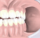 Teeth and gingiva
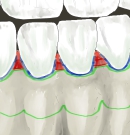 Parts of the gingiva
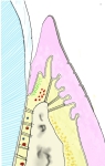 Gingiva (section)
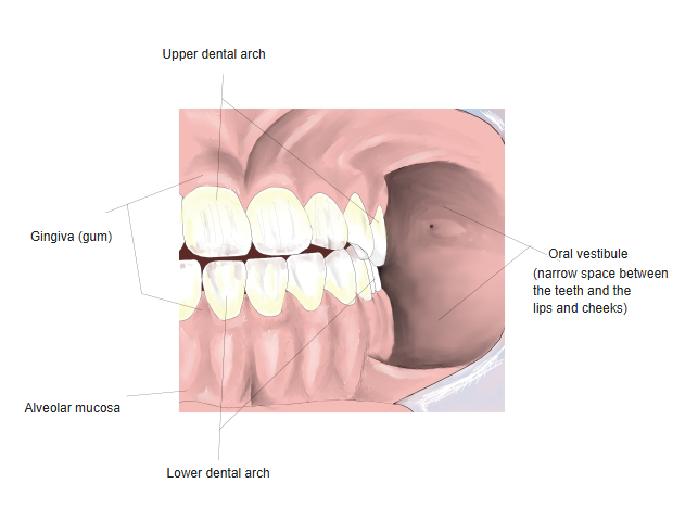
Reference websites
[Wikipedia]
· Gums, Wikipedia.
· Periodontium, Wikipedia.
[Websites with illustrations]
· Gingiva (figure) (Jaypee Digital)

· Gingiva (figure) (Springer Nature)

· Junctional epithelium (Periobasics)
Where it is found
Related terms
References and sources
- Further reading: Gums, Wikipedia.
- Further reading: Periodontium, Wikipedia.
- Federative International Programme for Anatomical Terminology (FIPAT). Terminologia Anatomica, 2nd ed. (TA2), 2019.