Gingival sulcus (Latin: sulcus gingivalis)
Also called Gingival crevice
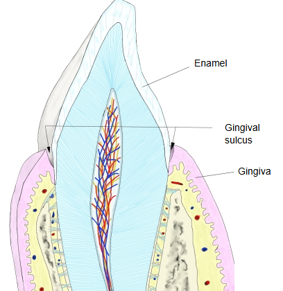
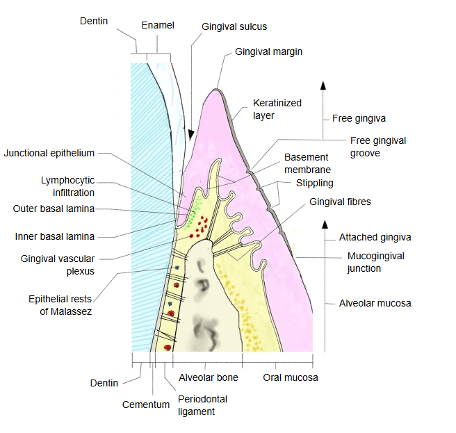
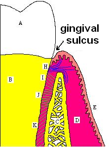
· It forms as the tooth erupts: the reduced enamel epithelium, which took part in making the enamel, partly separates from the enamel surface. Its floor is formed by the junctional epithelium, which attaches the gingiva to the tooth; its outer wall is the inner surface of the free gingiva.
· It is shallow around the front teeth and deeper around the premolars and molars.
· Its average depth is about 1.8 mm; clinically it measures about 0.5-2 mm. Sources differ on the normal depth (some give up to 3 mm), and clinical probing usually reads deeper than the histological depth.
· When dental plaque collects in the gingival sulcus, the bacteria in it release toxins and the gingiva becomes inflamed and swollen (gingivitis).
A sulcus that has become deeper because the gingiva is swollen by disease is called a gingival pocket or false pocket. One that has become deeper because the bottom of the pocket has moved towards the root (loss of attachment) is called a periodontal pocket or true pocket.
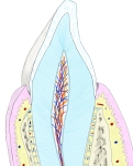 Tooth and gingiva (section)
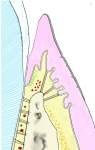 Free gingiva (section)
Reference websites
[Wikipedia]
· Gingival sulcus, Wikipedia.
· Gums, Wikipedia.
[Websites with illustrations]
· Gingival sulcus (diagram) (Wikimedia Commons)
· Parts of a human tooth (stock illustration) (Shutterstock)

· Gingiva and gingival sulcus (textbook figure) (Jaypee Digital)

Related terms
- Dental alveoli
- Alveolar border
- Alveolar canals
- Alveolar arch
- Alveolar foramina
- Alveolar bone
- Alveolar yokes
- Alveolar crest
- Apical foramen
- Gingiva
- Neck of tooth
- Periodontal ligament
- Crown
- Root of tooth
- Teeth
References and sources
- Further reading: Gingival sulcus, Wikipedia.
- Further reading: Gums, Wikipedia.
- Federative International Programme for Anatomical Terminology (FIPAT). Terminologia Anatomica, 2nd ed. (TA2), 2019.