Submental triangle (Latin: trigonum submentale)
Also called the suprahyoid triangle
Many of these contents in fact lie in the superficial fascia over the roof of the triangle rather than inside it.
It is one of the subdivisions of the anterior triangle of the neck, lying in the midline between the right and left submandibular triangles (see also carotid triangle), and it is the area of the submental region of the neck surface.
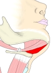 Submental triangle
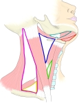 Triangles of the neck
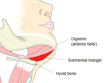
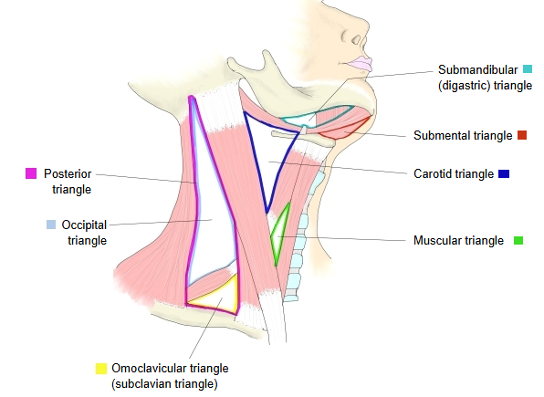
Below is a simple outline of the triangular depressions of the neck (it may not include every one).
depressions
of the neck
- Anterior triangle : sternocleidomastoid (anterior border), midline of the neck, body of mandible (lower border)
- Submandibular triangle : digastric (posterior and anterior bellies), body of mandible
- Submental triangle : anterior bellies of the left and right digastric, hyoid bone
- Carotid triangle : sternocleidomastoid, digastric (posterior belly), omohyoid (superior belly)
- Muscular triangle : sternocleidomastoid (anterior border), omohyoid, midline of the neck = omotracheal triangle
- Posterior triangle : sternocleidomastoid (posterior border), trapezius (anterior border), clavicle (middle third) = lateral cervical region
- Occipital triangle : trapezius (anterior border), sternocleidomastoid (posterior border), omohyoid (inferior belly)
- Omoclavicular triangle : omohyoid (inferior belly), sternocleidomastoid (posterior border), clavicle (middle third)
Reference websites
[Wikipedia]
· Submental triangle, Wikipedia.
[Websites with illustrations]
· Anterior triangle of the neck: boundaries, contents and subdivisions (TeachMeAnatomy)
· Anterior triangle of the neck (slide) (SlideShare)

· Submental triangle (figure) (AnatomyQA)

· Muscles of the neck with the triangles labelled (Wikimedia Commons)

Boundaries
The triangle is bounded as follows:
· Lateral (each side): the anterior belly of the digastric.
· Medial / apex: the midline of the neck between the mandible and the hyoid bone; the apex is at the chin, near the midline of the mandible. When the right and left halves are counted as one triangle, the two anterior bellies form its sides.
· Base (below): the body of the hyoid bone.
· Floor: the two mylohyoid muscles, which meet in the midline at the mylohyoid raphe.
· Roof: skin, superficial fascia (with the platysma at the sides) and the investing layer of the deep cervical fascia.
Contents
Submental lymph nodes (usually a few), which drain the chin, the middle of the lower lip, the floor of the mouth and the tip of the tongue; small submental veins that join to form the beginning of the anterior jugular vein; branches of the submental artery may run along its edge. Many of these structures lie in the superficial fascia over the roof rather than inside the triangle.
Related terms
- Carotid triangle
- Digastric
- Mylohyoid
- Body of hyoid bone
- Suprahyoid muscles
- Submental vein
- Anterior jugular vein
References and sources
- Further reading: Submental triangle, Wikipedia.
- Federative International Programme for Anatomical Terminology (FIPAT). Terminologia Anatomica, 2nd ed. (TA2), 2019.