Occipital triangle (Latin: trigonum occipitale)
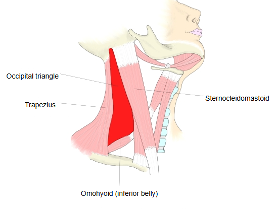
The occipital triangle is one of the triangular depressions seen in the neck; it is part of the posterior triangle.
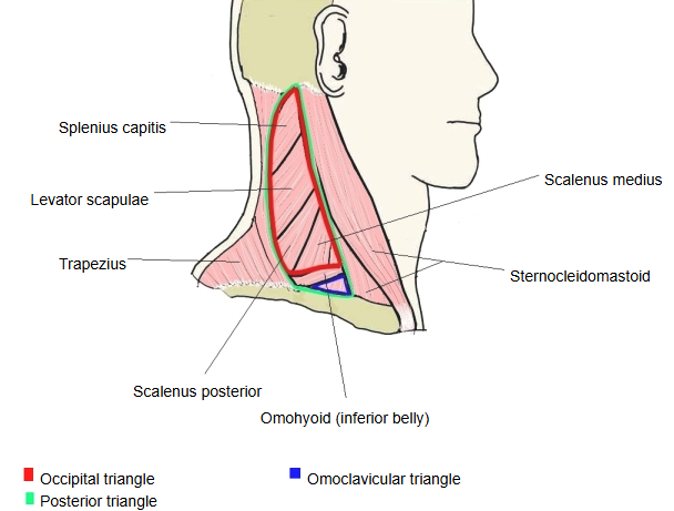
The floor is formed by splenius capitis, levator scapulae, and the scalenus medius and scalenus posterior.
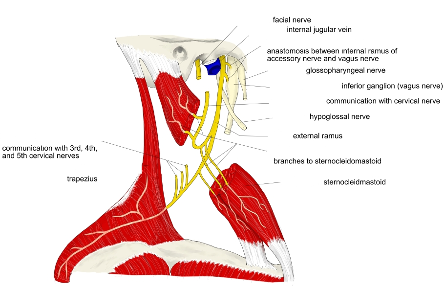
Main contents: external jugular vein, accessory nerve, cervical plexus (cutaneous branches) and brachial plexus (upper part).
Also the transverse cervical artery and lymph nodes along the posterior border of sternocleidomastoidCovered by skin, fascia and, in its lower part, the platysma
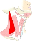 Occipital triangle
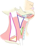 Triangles of the neck
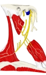 Accessory nerve
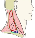 Posterior triangle and its parts
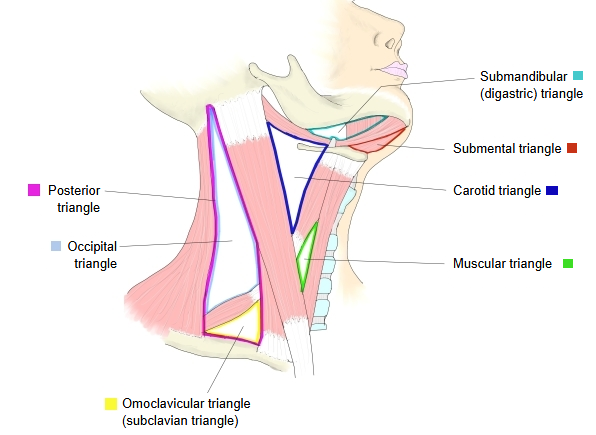
Below is a simple outline of the triangular depressions of the neck (it may not include every one).
Triangular
depressions
of the neck
depressions
of the neck
- Anterior triangle : sternocleidomastoid (anterior border), midline of the neck, body of mandible (lower border)
- Submandibular triangle : digastric (posterior and anterior bellies), body of mandible
- Submental triangle : anterior bellies of the left and right digastric, hyoid bone
- Carotid triangle : sternocleidomastoid, digastric (posterior belly), omohyoid (superior belly)
- Muscular triangle : sternocleidomastoid (anterior border), omohyoid, midline of the neck = omotracheal triangle
- Posterior triangle : sternocleidomastoid (posterior border), trapezius (anterior border), clavicle (middle third) = lateral cervical region
- Occipital triangle : trapezius (anterior border), sternocleidomastoid (posterior border), omohyoid (inferior belly)
- Omoclavicular triangle : omohyoid (inferior belly), sternocleidomastoid (posterior border), clavicle (middle third)
Reference websites
[Wikipedia]
· Occipital triangle, Wikipedia.
· Posterior triangle of the neck, Wikipedia.
[Websites with illustrations]
· Posterior triangle of the neck: subdivisions (TeachMeAnatomy)
· Muscles and labelled triangles of the neck (Wikimedia Commons)

Related terms
- Posterior triangle of neck
- Omoclavicular triangle
- Lateral cervical region
- Anterior triangle of neck
- Carotid triangle
- Cervical fascia
- Accessory nerve
References and sources
- Further reading: Occipital triangle, Wikipedia.
- Further reading: Posterior triangle of the neck, Wikipedia.
- Federative International Programme for Anatomical Terminology (FIPAT). Terminologia Anatomica, 2nd ed. (TA2), 2019.