Rectus sheath (Latin: vagina musculi recti abdominis)
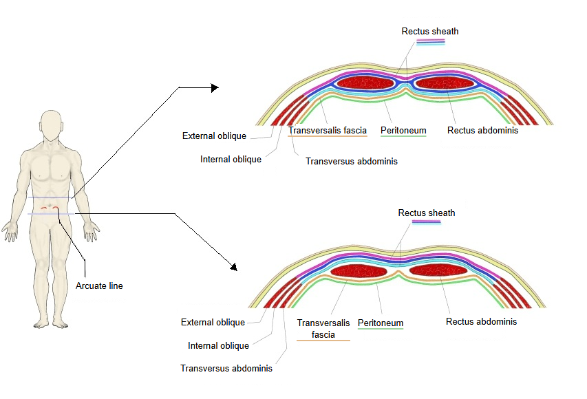
Above the arcuate line, the internal oblique aponeurosis splits at the lateral edge of the rectus into two laminae that pass in front of and behind the muscle. Below it, all three aponeuroses pass in front of the muscle, so behind the rectus lie only the transversalis fascia, a little extraperitoneal fat and the parietal peritoneum. Above the costal margin the internal oblique and transversus aponeuroses end, so only the external oblique aponeurosis covers the front and the muscle lies directly on the costal cartilages. Besides the rectus abdominis, the sheath contains the pyramidalis (when present), the superior and inferior epigastric vessels and the ends of the lower thoracic nerves. The aponeuroses of both sides meet and interlace in the midline to form the linea alba. Sources differ on details: some describe each aponeurosis as two-layered, and the level and sharpness of the arcuate line vary between people.
sheath
- Above the arcuate line
- Anterior layer : made of the aponeurosis of the external oblique and the aponeurosis of the internal oblique (its anterior lamina)
- Posterior layer : made of the aponeurosis of the internal oblique (its posterior lamina) and the aponeurosis of transversus abdominis
- Below the arcuate line
- Anterior layer : made of the aponeuroses of the internal and external obliques and of transversus abdominis
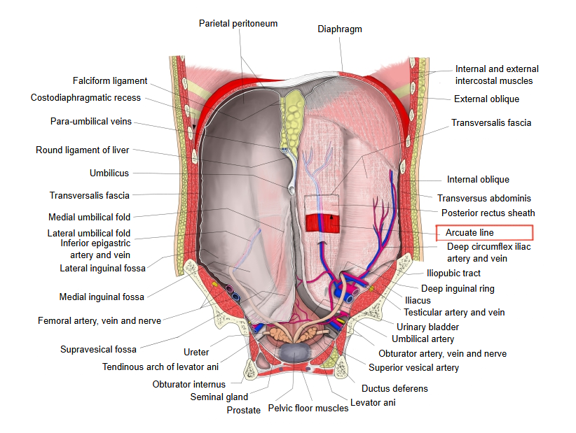
 Anterior abdominal wall from behind
Reference websites
[Wikipedia]
· Rectus sheath, Wikipedia.
· Arcuate line of rectus sheath, Wikipedia.
Related terms
- Rectus abdominis
- External oblique
- Internal oblique
- Transversus abdominis
- Transversalis fascia
- Parietal peritoneum
- Median umbilical ligament
References and sources
- Further reading: Rectus sheath, Wikipedia.
- Further reading: Arcuate line of rectus sheath, Wikipedia.
- Federative International Programme for Anatomical Terminology (FIPAT). Terminologia Anatomica, 2nd ed. (TA2), 2019.