Latin: os ilium
Overview
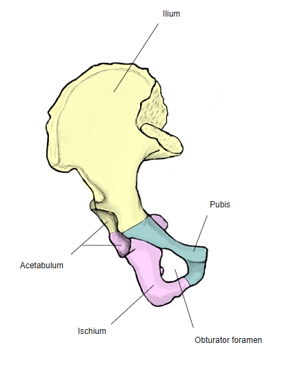
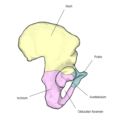
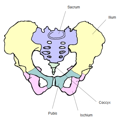
- One of the three bones that make up the hip bone (with the ischium and pubis) – the largest and uppermost of the three.
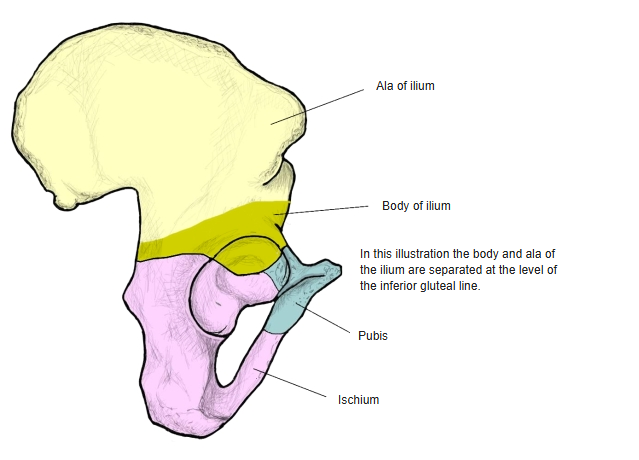
- It is divided into the body of the ilium, which forms the upper part (about two-fifths) of the acetabulum, and the broad, flat ala (wing) of the ilium that spreads upwards from it. The boundary between body and ala is not sharply defined; on the inner surface it lies roughly at the arcuate line.
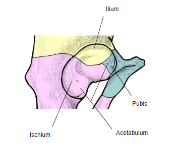
- Until puberty the ilium, ischium and pubis are joined by cartilage (the Y-shaped triradiate cartilage in the acetabulum); in adolescence they fuse by bone into a single hip bone.
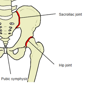
- Through the acetabulum the ilium takes part in the hip joint with the femur, and through its auricular surface it forms the sacroiliac joint with the sacrum.
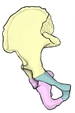
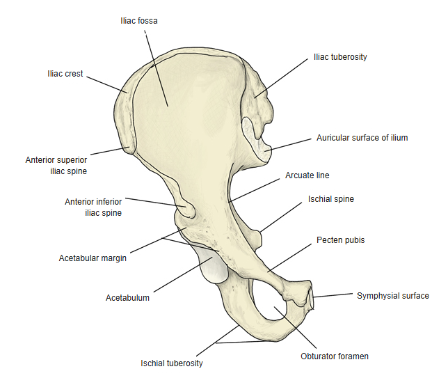
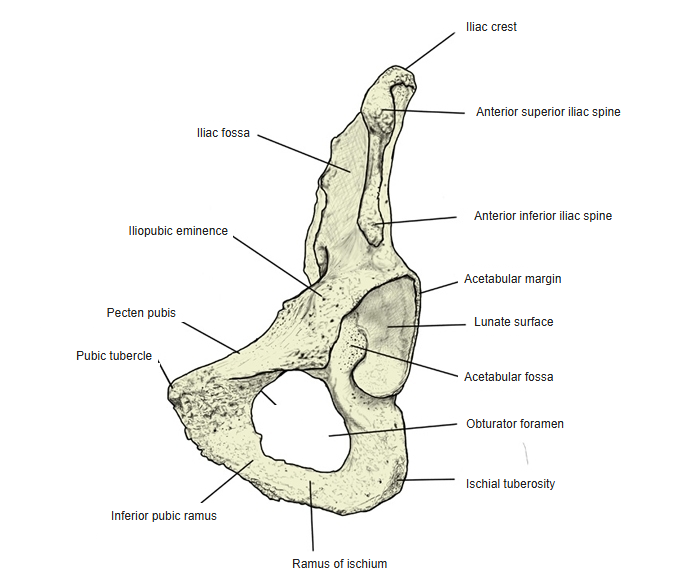
Right hip bone: ilium, ischium and pubis (lateral) 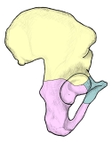
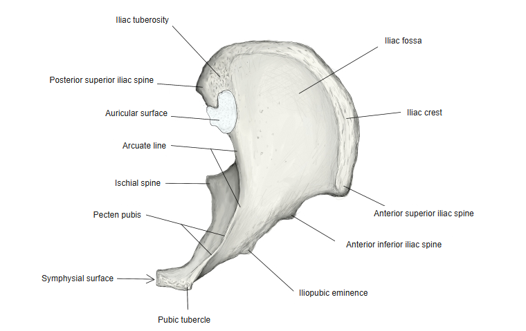
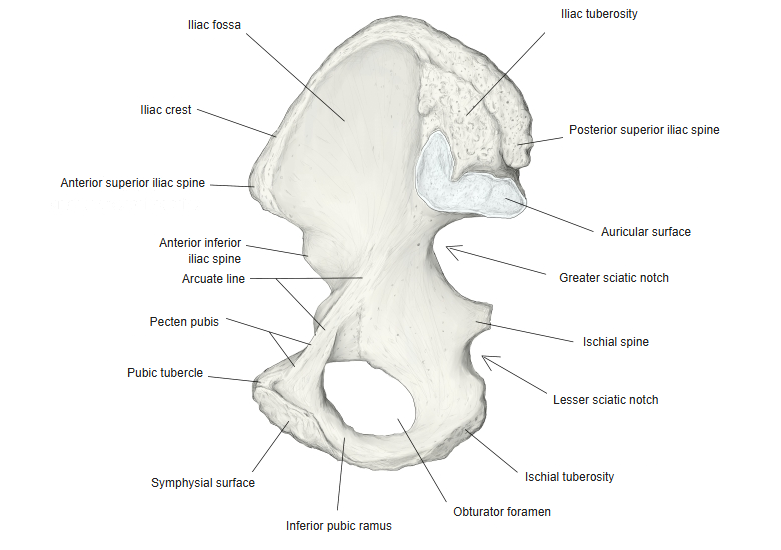
Right hip bone: ilium, ischium and pubis (medial)
- Bones of the lower limb (one side)
- Pelvic girdle
- Hip bone ← ilium, ischium and pubis fused
- Free lower limb
- Thigh and knee
- Femur
- Patella
- Leg
- Tibia ← big-toe side
- Fibula ← little-toe side
- Foot
- Tarsal bones (7)
- Metatarsal bones (5)
- Phalanges (14)
- Thigh and knee
- Pelvic girdle
Illustrations
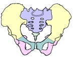
Pelvis: ilium, ischium, pubis, sacrum and coccyx 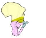
Body and ala of the ilium 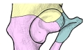
Acetabulum: contribution of the three bones 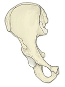
Right hip bone (anterior) 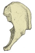
Left hip bone (from above) 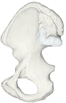
Right hip bone (medial) 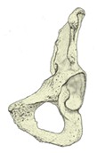
Right hip bone (anterior oblique)
Articulations
Ligaments
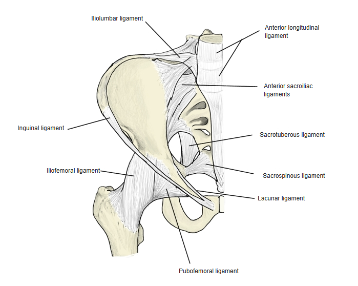
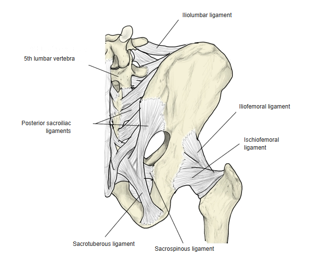
The following ligaments attach to the ilium. The list is intended to be comprehensive but may omit minor ligaments.
| # | Ligament | Connects |
|---|---|---|
| 1 | Anterior sacroiliac ligament | Front of the sacrum – front of the ilium |
| 2 | Interosseous sacroiliac ligament | Between the tuberosities of the sacrum and ilium |
| 3 | Posterior sacroiliac ligament (short and long) | Lateral sacral crest – posterior superior and inferior iliac spines |
| 4 | Sacrotuberous ligament | Ischial tuberosity – sacrum, coccyx and posterior inferior iliac spine |
| 5 | Iliolumbar ligament | Transverse process of L5 – iliac crest |
| 6 | Iliofemoral ligament | Anterior inferior iliac spine and acetabular rim – intertrochanteric line of the femur |
| 7 | Inguinal ligament | Anterior superior iliac spine – pubic tubercle |
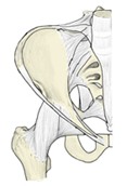
Ligaments of the pelvis (anterior) 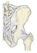
Ligaments of the pelvis (posterior)
Muscle attachments
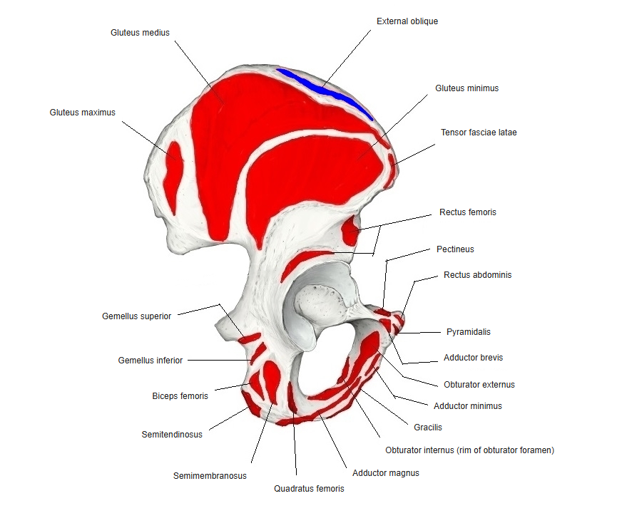
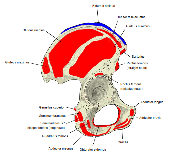
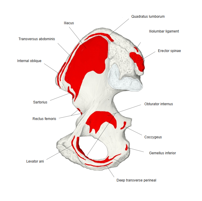
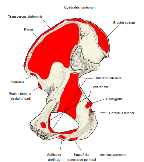
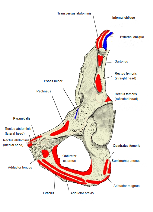
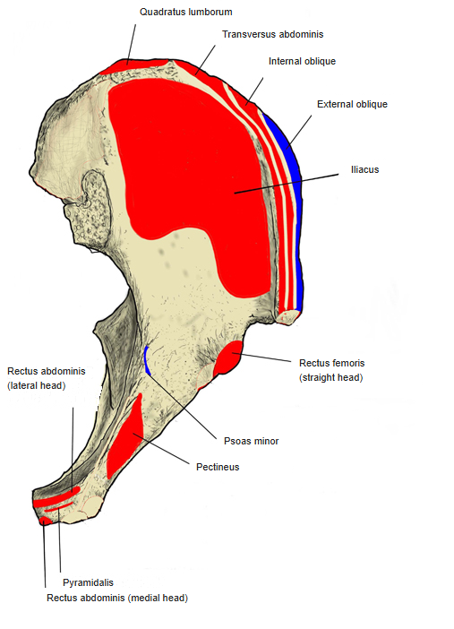
- Origin of: Latissimus dorsi, Internal oblique, Transversus abdominis, Quadratus lumborum, Iliocostalis lumborum, Gluteus maximus, Gluteus medius, Gluteus minimus, Iliacus, Tensor fasciae latae, Sartorius, Rectus femoris
- Insertion of: External oblique, Psoas minor
Iliac crest: latissimus dorsi, internal oblique, transversus abdominis, quadratus lumborum and iliocostalis lumborum arise, and external oblique inserts. Gluteal surface: gluteus maximus (behind the posterior gluteal line), medius and minimus. Iliac fossa: iliacus. Anterior superior iliac spine: sartorius and tensor fasciae latae. Anterior inferior iliac spine: rectus femoris. Psoas minor inserts near the iliopubic eminence.
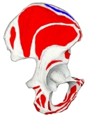
Muscle attachments (lateral surface) 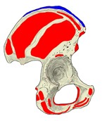
Muscle attachments (lateral surface) 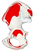
Muscle attachments (medial surface) 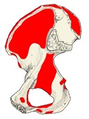
Muscle attachments (medial surface) 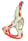
Muscle attachments (anterior oblique) 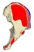
Muscle attachments (from above)
Parts
- Body of ilium – the lower, thick part; forms the upper part of the acetabulum.
- Ala of ilium – the broad upper part, with two surfaces (gluteal and sacropelvic) and four borders.
- Iliac crest – the thick, curved upper border; it has an outer lip, an intermediate zone and an inner lip, and a tubercle of the iliac crest on the outer lip about 5 cm behind the anterior superior iliac spine.
- Anterior superior iliac spine and anterior inferior iliac spine – the front end of the crest and the projection below it on the anterior border.
- Posterior superior iliac spine and posterior inferior iliac spine – the back end of the crest and the projection below it on the posterior border.
- Greater sciatic notch – the deep notch below the posterior inferior iliac spine (completed below by the ischium).
- Gluteal surface – the outer surface, marked by the posterior, anterior and inferior gluteal lines that separate the origins of the gluteal muscles.
- Iliac fossa – the smooth, concave front part of the inner (sacropelvic) surface; iliacus arises here.
- Arcuate line – the ridge below the iliac fossa that forms part of the pelvic brim.
- Auricular surface – the ear-shaped articular surface for the sacrum on the back of the inner surface; around it may lie the paraglenoid sulci.
- Iliac tuberosity – the rough area above and behind the auricular surface for the interosseous sacroiliac ligament.
- Iliopubic eminence – a low swelling where the ilium meets the pubis.
References and sources
- Further reading: Ilium (bone), Wikipedia.