Meridional fibres of ciliary muscle (Latin: fibrae meridionales musculi ciliaris)
Also called: Brücke's muscle, longitudinal fibres of the ciliary muscle
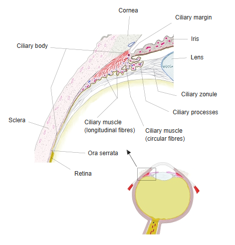
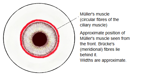
- Circular fibres : = Müller's muscle, the circular muscle in the front, inner part; how well it is developed varies greatly between individuals
- Longitudinal fibres : = Brücke's muscle, running meridionally along the junction of the cornea and the sclera to the ciliary body
- Radial fibres : between the circular and the longitudinal fibres; they fan out radially from the junction of the cornea and the sclera
They form the outer part of the ciliary muscle, just inside the sclera. They arise near the corneoscleral junction, from the posterior limiting lamina of the cornea, the scleral spur and the trabecular meshwork on the inner wall of the scleral venous sinus. They run backwards in a meridional direction, past the ciliary ring, and end in the stroma of the ciliary body and the suprachoroid layer of the choroid. Their contraction pulls the ciliary body and choroid forwards and inwards: this helps to relax the zonular fibres for accommodation and opens the trabecular meshwork, aiding the outflow of aqueous humour. Like the rest of the ciliary muscle they are supplied mainly by parasympathetic fibres from the oculomotor nerve.
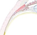 Ciliary body (section)
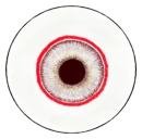 Müller's muscle (anterior view, diagram)
Reference websites
[Wikipedia]
· Ciliary muscle, Wikipedia.
[Websites with illustrations]
· The fibre groups of the ciliary muscle, showing the radial fibres (American Academy of Ophthalmology (image))
Related terms
References and sources
- Further reading: Ciliary muscle, Wikipedia.
- Federative International Programme for Anatomical Terminology (FIPAT). Terminologia Anatomica, 2nd ed. (TA2), 2019.