Zonular fibers (Latin: fibrae zonulares)
Also called the zonular fibres or the zonules
· They are suspending fibres attached to the equator and to the front and back surfaces of the lens near it; at their other end they attach to the basal lamina of the epithelium of the ciliary body.
· Each zonular fiber is only about 1–2 µm thick.
The fibers arise from the epithelium of the ciliary body, mainly from the flat ciliary ring and the valleys between the ciliary processes, and attach to the lens capsule in front of, at and behind the equator, all the way round. They are built mainly of fibrillin-rich microfibrils. Their tension keeps the lens flattened; when the ciliary muscle contracts, the tension falls and the lens becomes rounder for near vision.
 Ciliary zonule (diagram)
 Around the iris (section)
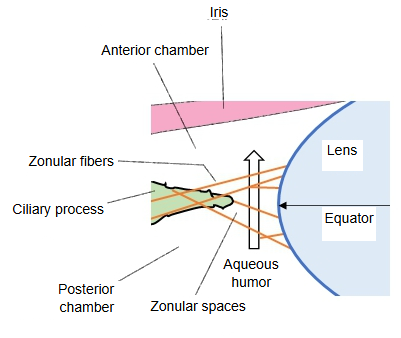
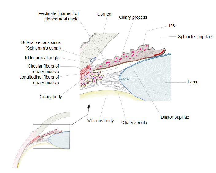
The zonular fibers anchor the equator of the lens and the neighbouring parts of its front and back surfaces to the ciliary body and the ciliary part of the retina. Parts of the fibers are probably made by the ciliary epithelial cells. Together the fibers form the ciliary zonule (zonule of Zinn), the ring of fibrous strands that links the ciliary body with the lens. The fibers are separated by the zonular spaces, which hold aqueous humor; behind the zonule these spaces form a ring-shaped canal round the equator of the lens (the canal of Petit).
Reference websites
[Wikipedia]
· Zonule of Zinn, Wikipedia.
[Websites with illustrations]
· Eye anatomy, sectional view (Blausen) (Wikimedia Commons)

· Zonules (figure) (Webvision)
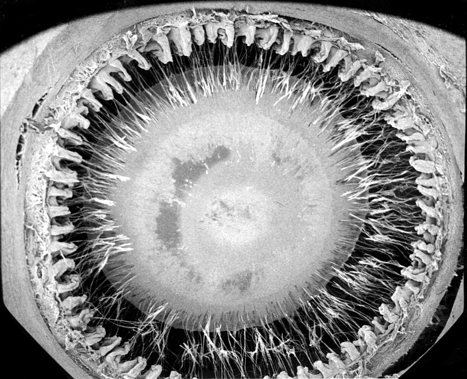
· Ciliary zonule and lens (figure) (entokey.com)
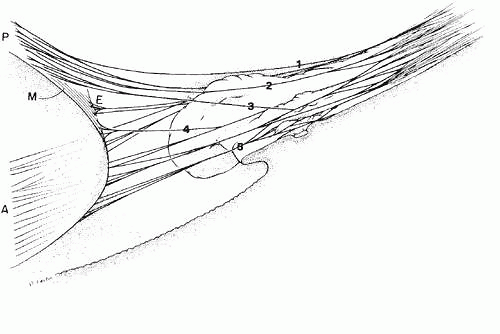
Related terms
References and sources
- Further reading: Zonule of Zinn, Wikipedia.
- Federative International Programme for Anatomical Terminology (FIPAT). Terminologia Anatomica, 2nd ed. (TA2), 2019.