Lateral process of malleus (Latin: processus lateralis mallei)
Also called the short process of the malleus.
Approximate sizes of the parts (the whole malleus is about 8–9 mm long):
1. 1. Handle of malleus: the longest part, a slender shaft running downwards.
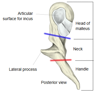
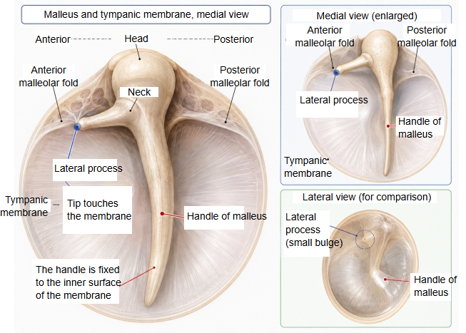
2. 2. Lateral process: very small, about 1 mm long or less; it projects straight laterally (towards the tympanic membrane) from where the neck meets the handle.
3. 3. Anterior process: a very thin, needle-like spur.
· The tip is fixed to the upper part of the tympanic membrane and pushes it outwards; seen from the ear canal this shows as a small bump, the malleolar prominence. The anterior and posterior malleolar folds run forwards and backwards from it and separate the small, slack pars flaccida above from the tense pars tensa below.
· Do not confuse it with the anterior process, a thin spur that runs forwards from the neck towards the petrotympanic fissure.
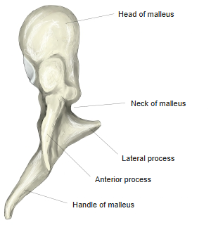
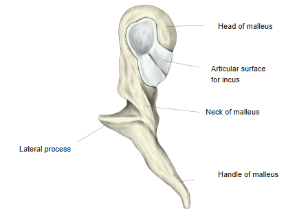
- Head of malleus : the rounded uppermost part, with the articular surface for the incus. Attachment of the superior ligament of the malleus.
- Neck of malleus : the narrow part just below the head; the two processes arise here or just below it (sources differ). Attachment of the lateral ligament of the malleus.
- Lateral process : projects laterally and presses into the tympanic membrane, forming the malleolar prominence.
- Anterior process : projects forwards and downwards; its tip reaches the petrotympanic fissure.
- Handle of malleus : long and slender, running downwards; it is the part attached to the tympanic membrane. The tendon of tensor tympani is attached near its upper end.
 Right tympanic cavity (coronal section)
 Right tympanic cavity (coronal section)
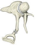 Left auditory ossicles (medial view)
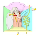 Right tympanic cavity (model, anterior view)
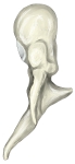 Malleus (anterior view)
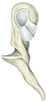 Malleus (posterior view)
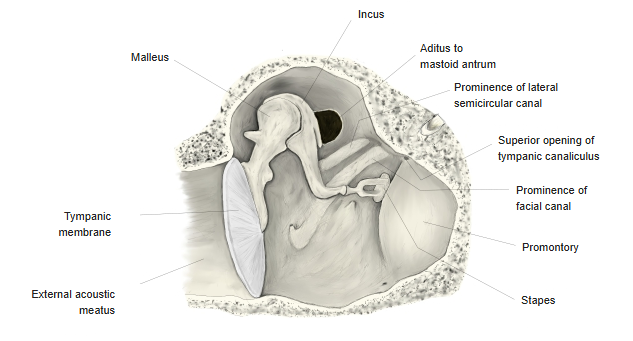
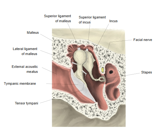
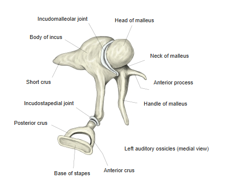
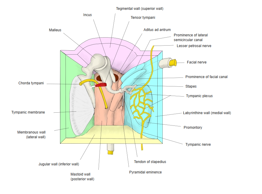
Reference websites
[Wikipedia]
· Malleus, Wikipedia.
[Websites with illustrations]
· Malleus (Gray's Anatomy, plate 916) (Wikimedia Commons)

· Malleus: anatomy and function (Kenhub)
· The middle ear: ossicles and their joints (TeachMeAnatomy)
Related terms
References and sources
- Further reading: Malleus, Wikipedia.
- Federative International Programme for Anatomical Terminology (FIPAT). Terminologia Anatomica, 2nd ed. (TA2), 2019.