Cochlear canaliculus (Latin: canaliculus cochleae)
Note: this page describes the cochlear canaliculus and the cochlear aqueduct separately, following one older account. Many points are still unclear, and most sources treat the two names as the same channel: many texts call the bony canal itself the cochlear aqueduct, while others keep that name for the perilymphatic duct inside it.
Simple definitions as used here:
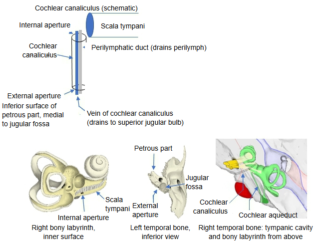
· Cochlear canaliculus: runs from the perilymphatic space to the external aperture of the cochlear canaliculus on the inferior surface of the petrous part of the temporal bone.
· Cochlear aqueduct: in that account, a separate channel running from the perilymphatic space medially toward the cranial cavity (in most sources, another name for the cochlear canaliculus).
Both carry the perilymphatic duct and a vein.
The cochlear canaliculus can be defined as follows.
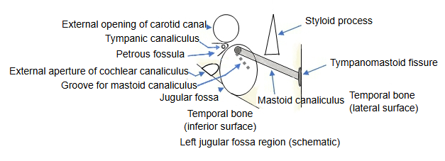
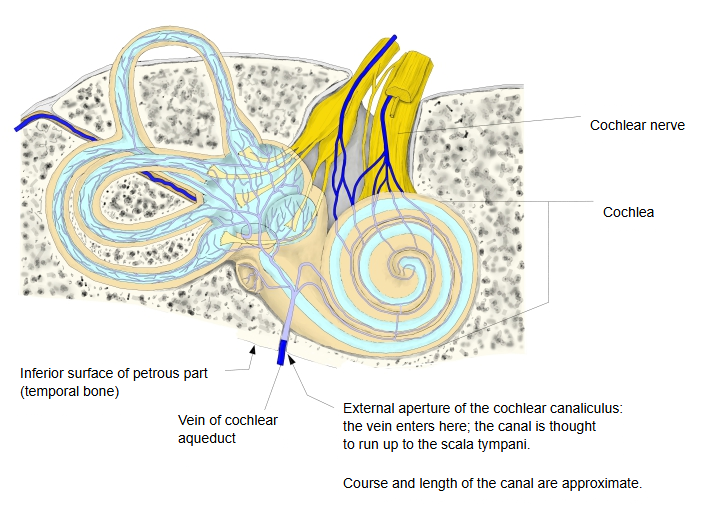
It is the passage of the perilymphatic duct of the internal ear: it begins in the scala tympani of the basal turn of the cochlea, close to the round window, and reaches the external aperture on the inferior surface of the petrous part. The external aperture is a small opening in front of and medial to the jugular fossa, behind and medial to the opening of the carotid canal.
Note: it is probably a short canal running roughly up and down, but few sources describe it, and its exact course and length are uncertain.
Reference websites
[Wikipedia]
· Cochlear aqueduct, Wikipedia.
· Perilymphatic duct, Wikipedia.
[Websites with illustrations]
· Interior of the right osseous labyrinth (Gray's Anatomy, fig. 921) (Wikimedia Commons)

· Base of the skull, external surface (Gray's Anatomy, fig. 187) (Wikimedia Commons)

· The inner ear: bony and membranous labyrinth (TeachMeAnatomy)
Related terms
- Perilymphatic duct
- Perilymphatic space
- Vein of cochlear aqueduct
- Petrous part
- Jugular foramen
- Cochlea
- Bony labyrinth
References and sources
- Further reading: Cochlear aqueduct, Wikipedia.
- Further reading: Perilymphatic duct, Wikipedia.
- Federative International Programme for Anatomical Terminology (FIPAT). Terminologia Anatomica, 2nd ed. (TA2), 2019.