Orbital fat body (Latin: corpus adiposum orbitae)
Also called the orbital fat or the retrobulbar fat
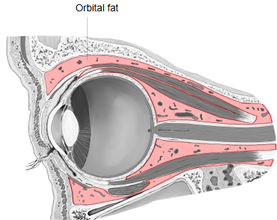
· It fills the spaces of the orbit around and behind the eyeball, packing the gaps between the eyeball, the extra-ocular muscles, the optic nerve, the vessels (ophthalmic artery and its branches) and nerves of the orbit and the lacrimal gland.
· It is divided by fine connective-tissue septa into small lobules; the sheath of the eyeball (fascial sheath, Tenon's capsule) separates it from the eyeball itself.
· It is often described as two parts: a central (intraconal) part inside the cone of the rectus muscles, and a peripheral (extraconal) part between the muscles and the bony walls.
· It cushions and supports the eyeball and lets it and the muscles move smoothly. Loss of this fat makes the eye look sunken; swelling of it pushes the eye forwards. See also accessory structures of the eye.
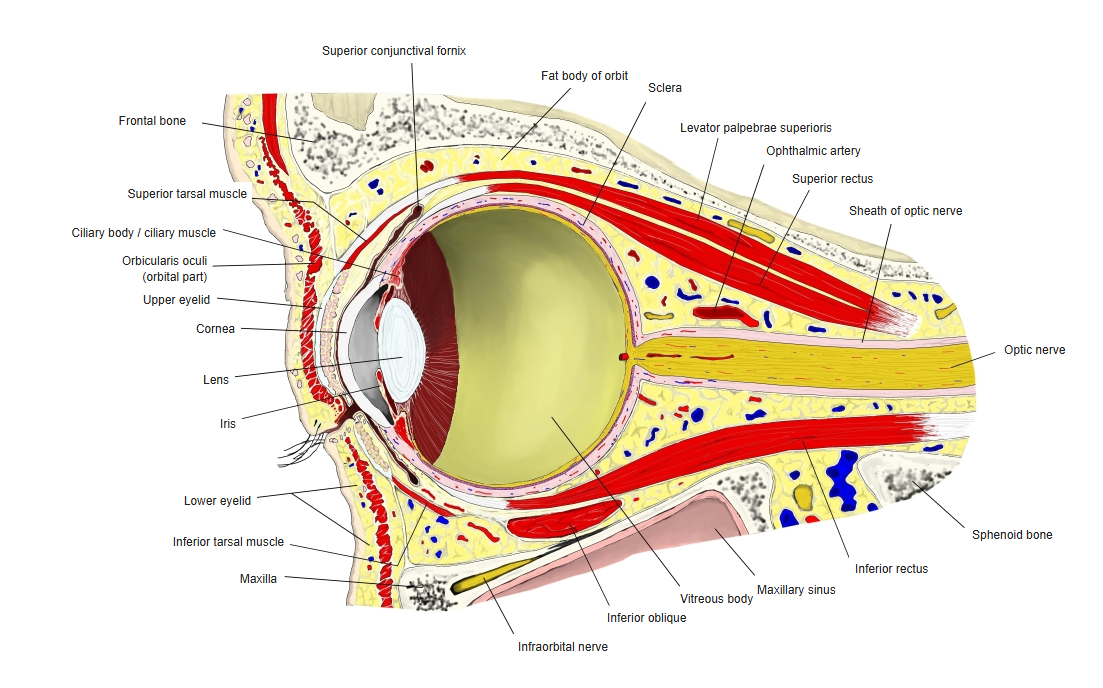
- Eyeball : about 24 mm from front to back. Main parts: cornea, anterior chamber, lens, vitreous body.
- Muscles : intrinsic muscles of the eyeball (ciliary muscle and others); extra-ocular muscles (superior rectus and others); others (levator palpebrae superioris and others).
- Vessels : arteries: ophthalmic artery, supratrochlear artery, lacrimal artery and others; veins: superior ophthalmic vein, lacrimal vein and others.
- Ligaments : Lockwood's ligament, Whitnall's ligament and others.
- Nerves : optic nerve, nasociliary nerve, lacrimal nerve and others.
- Others : lacrimal apparatus (lacrimal gland, lacrimal sac and others) and fat (the orbital fat body).
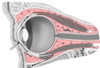 Orbital fat
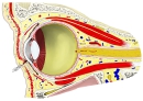 Orbit, sagittal section
Related terms
References and sources
- Further reading: Orbital fat, Wikipedia.
- Further reading: Orbit (anatomy), Wikipedia.
- Federative International Programme for Anatomical Terminology (FIPAT). Terminologia Anatomica, 2nd ed. (TA2), 2019.