Introitus of facial canal (Latin: introitus canalis facialis)
Also called the entrance of facial canal or the inner opening of facial canal
· The introitus is the inner opening of the facial canal; the outer opening is the stylomastoid foramen, which opens behind the root of the styloid process.
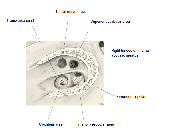
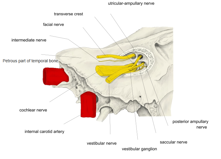
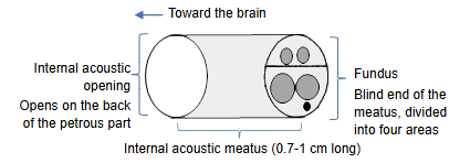
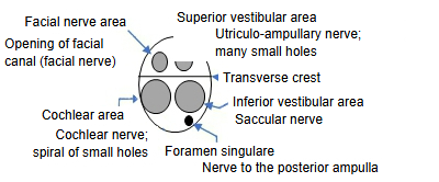
It lies in the upper front part of the fundus, above the transverse crest and in front of the superior vestibular area. The facial nerve passes through it together with the nervus intermedius. The first part of the canal, just beyond the introitus, is its narrowest part; this is why swelling of the facial nerve can compress it here.
- Upper part
- Front: facial nerve area : has one large opening, the introitus of the facial canal, through which the facial nerve passes.
- Back: superior vestibular area : many small holes for the utriculo-ampullary nerve, to the macula of the utricle and the ampullae of the anterior and lateral semicircular ducts.
- Lower part
- Front: cochlear area : many small holes in a spiral, for the cochlear nerve.
- Back: inferior vestibular area : small holes for the saccular nerve to the macula of the saccule. Behind and below it is a single hole, the foramen singulare, for the posterior ampullary nerve to the ampulla of the posterior semicircular duct.
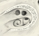 Fundus of the right internal acoustic meatus
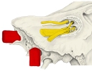 Nerves at the fundus
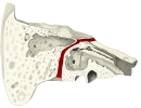 Facial canal (oblique section)
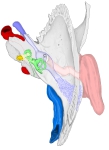 Right temporal bone and ear (superior view)
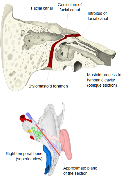
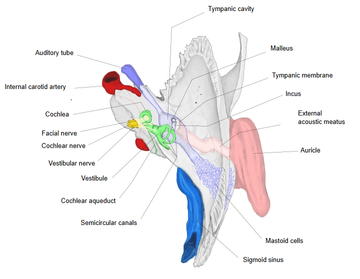
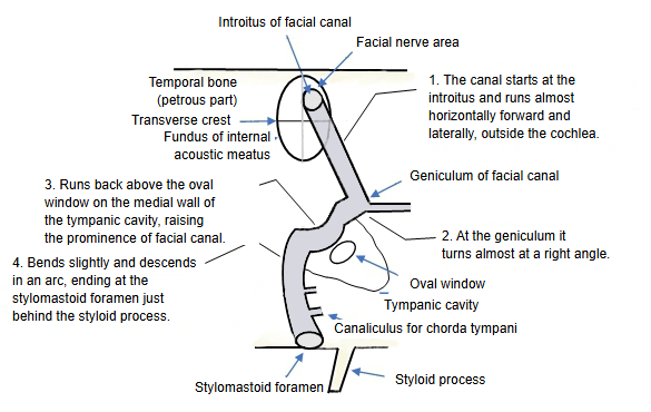
From the introitus the facial canal runs through the petrous part of the temporal bone, almost horizontally forward and laterally above the cochlea. At the geniculum of the facial canal, where the geniculate ganglion lies, it turns back almost at a right angle. It then runs backward on the medial wall of the tympanic cavity, above the oval window, where it raises the prominence of the facial canal. Behind the tympanic cavity it bends and descends in an arc to end at the stylomastoid foramen.
Reference websites
[Wikipedia]
· Facial canal, Wikipedia.
· Internal auditory meatus, Wikipedia.
[Websites with illustrations]
· Internal acoustic meatus: anatomy and function (Kenhub)
· The facial nerve (CN VII) (TeachMeAnatomy)
Related terms
- Internal acoustic meatus
- Transverse crest
- Stylomastoid foramen
- Facial nerve
- Geniculate ganglion
- Petrous part
- Temporal bone
References and sources
- Further reading: Facial canal, Wikipedia.
- Further reading: Internal auditory meatus, Wikipedia.
- Federative International Programme for Anatomical Terminology (FIPAT). Terminologia Anatomica, 2nd ed. (TA2), 2019.