Transverse crest (Latin: crista transversa)
Also called the falciform crest
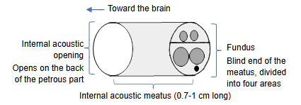
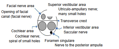
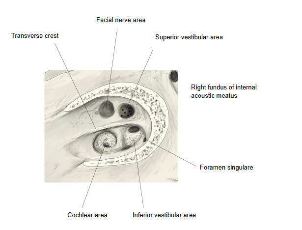
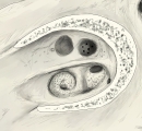 Right fundus of the internal acoustic meatus
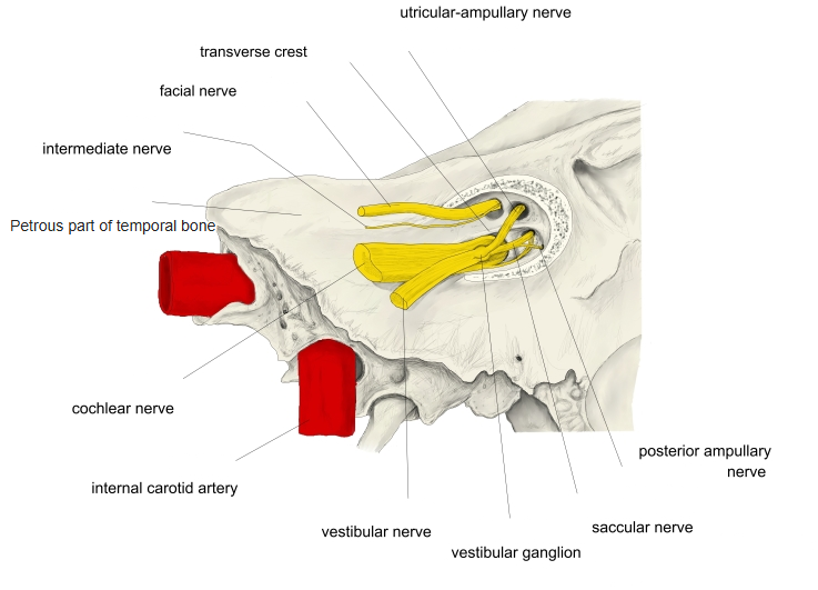
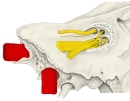 Fundus and nerves
- Squamous part : the part that takes up the middle and the whole upper part of the bone; it has a temporal surface and a cerebral surface.
- Petrous part
- Mastoid part : below the squamous part (from the mastoid process to the parietal notch).
- Pyramid : a three-sided pyramid running forwards and medially from the boundary of the squamous and mastoid parts; it has an apex, surfaces and borders.
- Apex : points towards the foramen lacerum; the internal opening of the carotid canal opens here.
- Anterior surface : = cerebral surface: trigeminal impression, arcuate eminence, grooves for the greater and lesser petrosal nerves.
- Posterior surface : = cerebellar surface: internal acoustic opening (leading to the meatus with the transverse crest at its end), subarcuate fossa, external aperture of the vestibular aqueduct.
- Inferior surface : = basal surface: styloid process, jugular fossa, groove for the mastoid canaliculus, external aperture of the cochlear canaliculus, external opening of the carotid canal, petrous fossula, inferior opening of the tympanic canaliculus.
- Tympanic part : the smallest part: a thin, half-tube-shaped plate of bone that forms the front and lower walls of the external acoustic meatus.
The internal acoustic meatus opens on the posterior surface of the petrous part and ends blindly at the fundus, a thin vertical plate of bone facing the inner ear. The transverse crest (also called the falciform crest, crista falciformis) runs horizontally across this plate and divides it into two unequal parts.
Above the crest lie the facial nerve area (front) and the superior vestibular area (back), separated by a small vertical ridge often called Bill's bar.
Below the crest lie the cochlear area (front, a spiral of small holes) and the inferior vestibular area (back), with the separate foramen singulare behind them.
The crest is a landmark for the nerves entering the inner ear: the facial nerve and the superior vestibular nerve run above it, the cochlear nerve and the inferior vestibular nerve below it.
Reference websites
[Wikipedia]
· Internal auditory meatus, Wikipedia.
[Websites with illustrations]
· Fundus of the right internal acoustic meatus, diagram (Gray's Anatomy, fig. 140) (Wikimedia Commons)

· Left temporal bone, inner surface (Gray's Anatomy, fig. 138) (Wikimedia Commons)

Related terms
- Internal acoustic meatus
- Petrous part
- Temporal bone
- Facial nerve
- Vestibulocochlear nerve
- Cochlear nerve
- Internal ear
References and sources
- Further reading: Internal auditory meatus, Wikipedia.
- Federative International Programme for Anatomical Terminology (FIPAT). Terminologia Anatomica, 2nd ed. (TA2), 2019.