Internal acoustic meatus (Latin: meatus acusticus internus)
Also called the internal auditory meatus or the internal auditory canal
Tumours of the nerve sheath of the vestibulocochlear nerve (vestibular schwannomas, also called acoustic neuromas) often start in the canal and widen it; they cause hearing loss and tinnitus on one side.
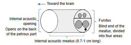
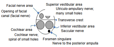
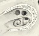 Fundus of the right internal acoustic meatus
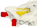 Fundus of the internal acoustic meatus and the nerves
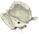 Left temporal bone (medial surface)
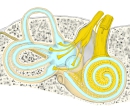 Right ear (front view)
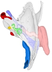 Right temporal bone (superior view)
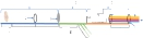 Hearing: model diagram
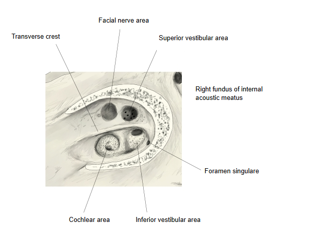
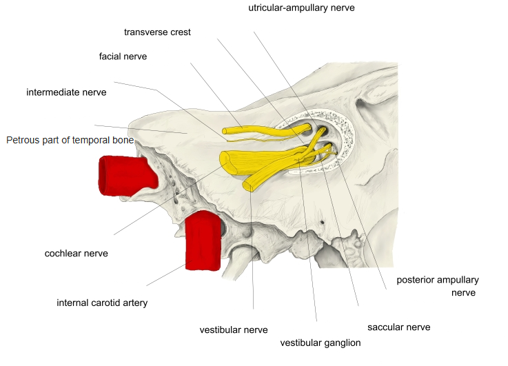
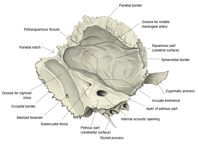
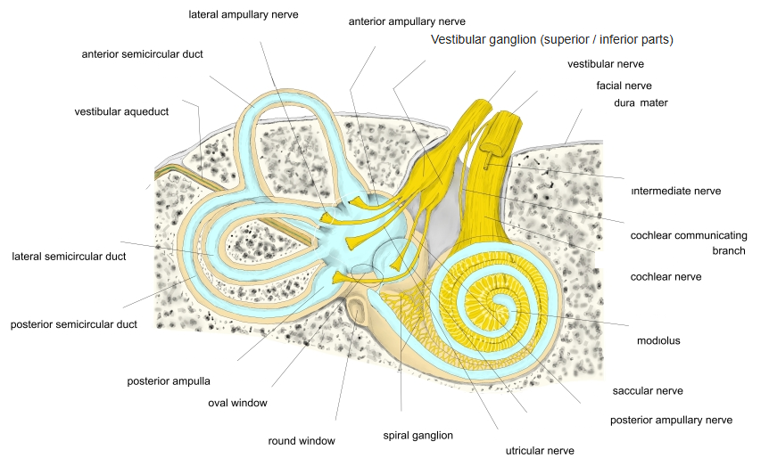
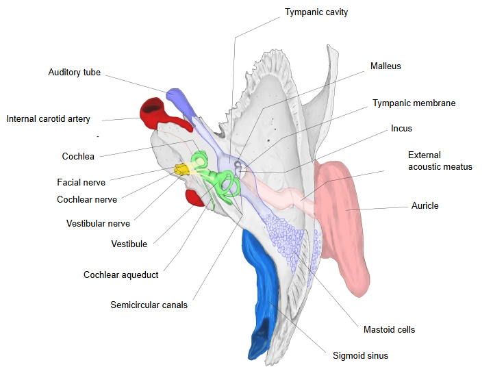
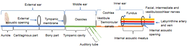
The internal acoustic meatus is the passage of the facial nerve, the intermediate nerve, the vestibulocochlear nerve and the labyrinthine artery and veins. Its blind end, the fundus, is divided by the transverse crest into an upper and a lower part, and both parts have many small holes. The somewhat larger hole at the front of the upper part is the beginning of the facial canal; the other small holes carry the branches of the vestibulocochlear nerve.
[Fundus]
- Upper part
- Facial nerve area : front: the opening of the facial canal, for the facial and intermediate nerves.
- Superior vestibular area : back: holes for the nerve fibres to the utricle and the ampullae of the anterior and lateral semicircular ducts.
- Lower part
- Cochlear area : front: a spiral of small holes for the cochlear nerve.
- Inferior vestibular area : back: holes for the nerve fibres to the saccule.
- Foramen singulare : a single hole behind the inferior vestibular area, for the nerve to the ampulla of the posterior semicircular duct.
The upper and lower parts are separated by the horizontal transverse crest (falciform crest); a small vertical ridge (often called Bill's bar) separates the two areas of the upper part. So the fundus has four areas, and the foramen singulare is a fifth, separate opening. A common way to remember the layout: the facial nerve lies upper front and the cochlear nerve lower front.
Reference websites
[Wikipedia]
· Internal auditory meatus, Wikipedia.
[Websites with illustrations]
· Internal acoustic meatus: anatomy and function (Kenhub)
· The inner ear (TeachMeAnatomy)
· The cochlea and vestibule viewed from above (Gray's Anatomy, fig. 923) (Wikimedia Commons)

Where it is found
Related terms
- Vestibulocochlear nerve
- Facial nerve
- Labyrinthine veins
- Petrous part
- External acoustic meatus
- Cochlea
- Ear (vestibulocochlear organ)
References and sources
- Further reading: Internal auditory meatus, Wikipedia.
- Federative International Programme for Anatomical Terminology (FIPAT). Terminologia Anatomica, 2nd ed. (TA2), 2019.