Lamina cribrosa of sclera (Latin: lamina cribrosa sclerae)
Also called Lamina cribrosa or the cribriform plate of sclera
· Some sources call it the cribriform plate of the sclera.
In more detail:
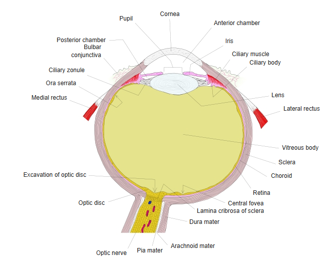
It is the part where the optic nerve, coming from the retina, pierces the sclera near the posterior pole of the eye, on its nasal side. Here the sclera becomes a sieve-like mesh of layers of collagen fibres stretched across the scleral canal, and the axons of the retinal ganglion cells pass through its many small pores in bundles. The central retinal artery and central retinal vein pass through near its centre. It is thinner and weaker than the rest of the sclera, so it is pushed backward when the pressure inside the eye rises; this bowing can distort the pores and damage the nerve fibres, and is thought to be one cause of nerve damage in glaucoma. Behind the eye the optic nerve runs to the optic canal.
Reference websites
[Wikipedia]
· Lamina cribrosa sclerae, Wikipedia.
· Glaucoma, Wikipedia.
[Websites with illustrations]
· Regions of the optic nerve head, with the lamina cribrosa region (figure) (Semantic Scholar)

· Lamina cribrosa with the central retinal vessels (figure) (Elsevier)

· Optic nerve head and lamina cribrosa (figure) (American Academy of Ophthalmology)
Related terms
References and sources
- Further reading: Lamina cribrosa of the sclera, Wikipedia.
- Federative International Programme for Anatomical Terminology (FIPAT). Terminologia Anatomica, 2nd ed. (TA2), 2019.