Ora serrata (Latin: ora serrata)
· Towards the margin of the optic part of the retina, the number of rod cells increases.
· The name describes an edge with fine saw-like teeth.
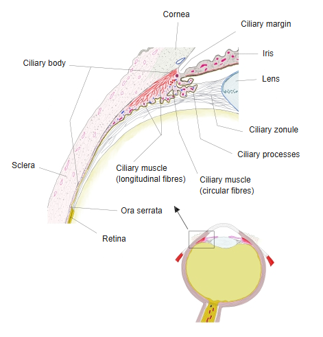
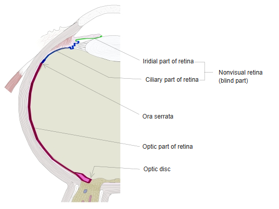
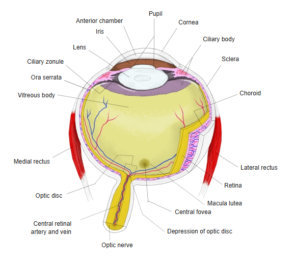
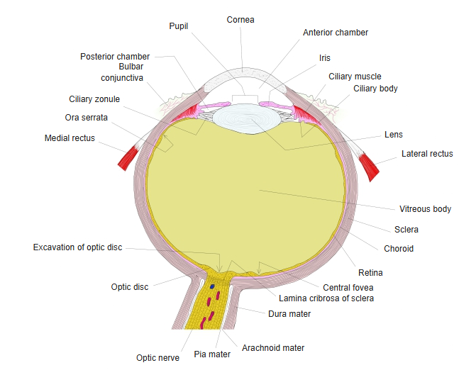
The ora serrata runs all the way round the inside of the eyeball, a few millimetres behind the corneoscleral junction. Here the multilayered neural retina stops; only its two-layered epithelium continues forwards over the ciliary body and the back of the iris. The retina is very thin here and firmly attached to the layers beneath it (choroid), so a retinal detachment usually stops at the ora serrata. The retina forms the inner layer of the eyeball, the innermost of the three layers of the wall of the eyeball.
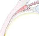 Around the ciliary body (horizontal section)
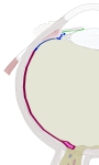 Optic part and blind part of the retina
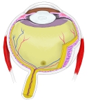 Eyeball (horizontal section)
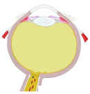 Right eyeball (horizontal section, schematic)
Older texts describe it in more detail:
The ora serrata reaches somewhat further forward on the medial (nasal) side than on the lateral (temporal) side; it is not centred, so the ciliary ring is wider laterally than medially. In development the number of its teeth matches the number of the large processes of the ciliary body (about 70), but many are lost later. Its form varies greatly: the teeth may be barely marked, and on the lateral side of the eyeball they may be missing altogether, while on the medial side they are always best developed. So not only the number of teeth but the whole shape of the edge of the retina varies considerably here.
Reference websites
[Wikipedia]
· Ora serrata, Wikipedia.
[Websites with illustrations]
· Schematic diagram of the human eye (Wikimedia Commons)

Related terms
References and sources
- Further reading: Ora serrata, Wikipedia.
- Federative International Programme for Anatomical Terminology (FIPAT). Terminologia Anatomica, 2nd ed. (TA2), 2019.