Optic part of retina (Latin: Pars optica retinae)
Also called the visual retina
- Overview
- Cells
- Layers
- Pathway
- Special regions
- Related terms
- Reference websites
- (Wikipedia, websites with illustrations)
- Related terms
Overview
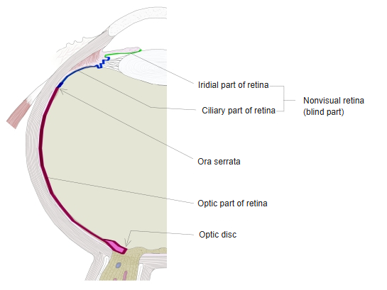
· In contrast to the optic part, the front part of the retina (the ciliary part and the iridial part), which cannot sense light, is called the blind part of the retina (non-visual retina).
· In life it is transparent like glass and smooth; after death it becomes cloudy and folds easily. Light passes through its inner layers before it reaches the photoreceptors, which lie on the outer side (an 'inverted' retina).
of the
retina
- Optic part of the retina : the part that senses light; made of nerve cells and connective (supporting) tissue, in which 10 layers can be distinguished.
- Blind part of the retina : cannot sense light; begins in front of the ora serrata.
- Ciliary part of the retina : a single layer of epithelial cells rich in pigment.
- Iridial part of the retina : a single layer of epithelial cells (without pigment but containing fine granules).
 Parts of the retina
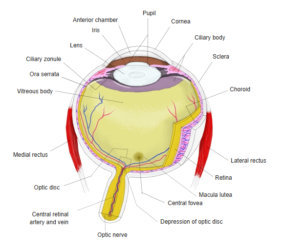
 Eyeball (horizontal section)
Cells
The cells of the optic part of the retina fall into two groups: the nerve cells that pass on information, and the cells that make up the supporting (connective) tissue.
optic part
of the retina
- Nerve cells
- Photoreceptor cells : rod cells (the majority) and cone cells
- Others : horizontal cells, bipolar cells, amacrine cells, ganglion cells
- Supporting cells (a kind of glial cell)
- Müller cells : span the retina like pillars
- Neuroglia : fill the spaces between the nerve cells and support them
of the
optic part
of the
retina
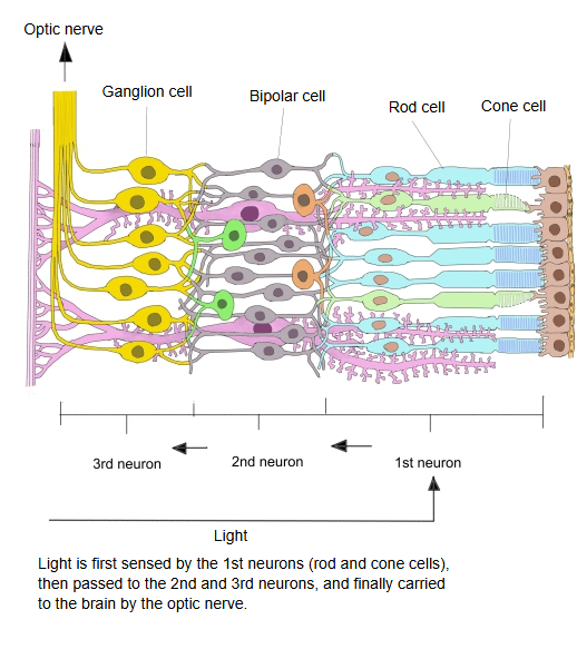
- Photoreceptor cells
- Rod cell : rod-shaped outer segment; most photoreceptors are rods. They serve light reception in dim light.
- Cone cell : cone-shaped outer segment; concentrated in the centre of the retina. They work in bright light and give sharp and colour vision.
- Horizontal cell : its processes spread sideways along the retina and synapse with the photoreceptor cells.
- Bipolar cell : has two processes, one on each side of the cell body; passes the signal from the photoreceptors to the ganglion cells.
- Amacrine cell : a local interneuron without a long axon; links bipolar cells and ganglion cells.
- Ganglion cell : has a long axon that runs to the brain; the axons form the optic nerve.
- Müller cell : not a neuron but a kind of glial cell; it spans the full thickness of the retina as a supporting cell and does not carry the visual signal.
Layers
From outside inwards, the optic part of the retina is usually described as having the following 10 layers.
| Layer | Description | |
|---|---|---|
| 1 | Layer of rods and cones | Formed by the outer segments of the photoreceptor cells (rod cells and cone cells), that is, the rod- and cone-shaped projections. |
| 2 | Outer limiting membrane | Like the inner limiting membrane, formed by the processes of the Müller cells. |
| 3 | Outer nuclear (granular) layer | The cell bodies of the rod cells and cone cells. Each has a nucleus and two processes: one runs as a rod or cone fibre into the outer plexiform layer, where it meets the processes of the bipolar cells of the inner nuclear layer; the other runs the opposite way as the rod or cone itself into the layer of rods and cones. |
| 4 | Fibre layer of Henle | The obliquely running rod and cone fibres. Some texts count it as part of the outer plexiform layer, not as a separate layer; sources differ. |
| 5 | Outer plexiform layer | Mainly the processes of the rod and cone cells and the dendrites of the bipolar cells, where the photoreceptors synapse with the bipolar and horizontal cells; it also contains fibres of the horizontal cells, amacrine cells and Müller cells. |
| 6 | Inner nuclear (granular) layer | Several rows of bipolar cells, whose processes branch in the inner and outer plexiform layers; it also holds the cell bodies of the amacrine, horizontal and Müller cells. · Bipolar cells: each sends one process outwards to the outer plexiform layer, where it synapses with photoreceptor cells, and one inwards to the inner plexiform layer, where it synapses with the ganglion cells (multipolar neurons). · Amacrine cells: cells with short processes in the innermost part of the layer, which synapse with the bipolar cells. · Horizontal cells: in the outer part of the layer; their processes synapse with the photoreceptor cells and are thought to link photoreceptors with each other. · Müller cells: a special kind of glial cell, long cells that run radially through the retina from the outer to the inner limiting membrane. |
| 7 | Inner plexiform layer | The axons of the bipolar cells of the inner nuclear layer, the processes of the amacrine cells and Müller cells, and the dendrites of the ganglion cells; here the bipolar and amacrine cells synapse with the ganglion cells. |
| 8 | Ganglion cell layer | A single layer of multipolar neurons (the ganglion cells). Their axons make up most of the nerve fibre layer, and their dendrites take part in the inner plexiform layer. |
| 9 | Nerve fibre layer | The axons of the ganglion cells, which run to the optic disc and form the optic nerve to the brain. |
| 10 | Inner limiting membrane | Formed by the end-feet of the Müller cells. Neurons of the retina: in the optic part three neurons are distinguished, the photoreceptor cells, the bipolar cells and the ganglion cells (multipolar neurons). |
Pathway
A light stimulus reaching the optic part of the retina is first sensed by the first neurons, the rod cells and cone cells, which lie in the outermost layer of the retina,
and is passed from them to the second neurons and then to the third neurons.
The path of the visual signal in the retina
Special regions
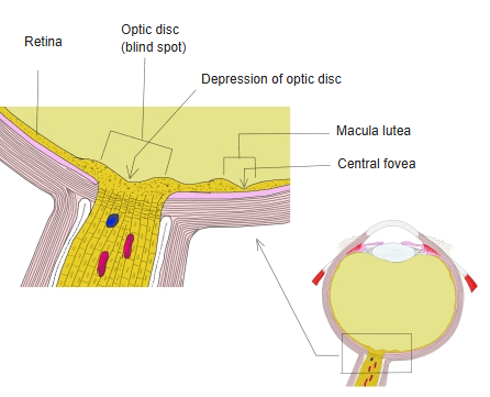
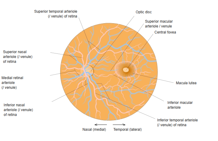
Three parts of the retina are usually described as special regions:
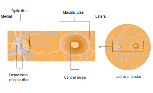
1. 1. Optic disc: the oval area, seen from inside the eyeball, where the optic nerve leaves the eye; it has no photoreceptors (the blind spot).
2. 2. Macula lutea: lateral to the optic disc, with the central fovea, the part of sharpest vision.
3. 3. Ora serrata: the very thin, scalloped border between the optic part of the retina and the blind part of the retina.
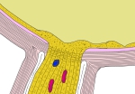 Optic disc and macula (section)
- Eyeball (horizontal section)
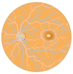 Fundus of the eye
 Optic disc and macula lutea
Related terms
Blind spot (Mariotte's blind spot), depression of optic disc (= cup of the optic disc), fovea centralis, macula lutea, Müller cell, neuroglia, optic disc (= optic papilla), ora serrata.
Reference websites
[Wikipedia]
· Retina, Wikipedia.
· Photoreceptor cell, Wikipedia.
[Websites with illustrations]
· Parts of the eye, with the retina (diagram) (National Eye Institute)
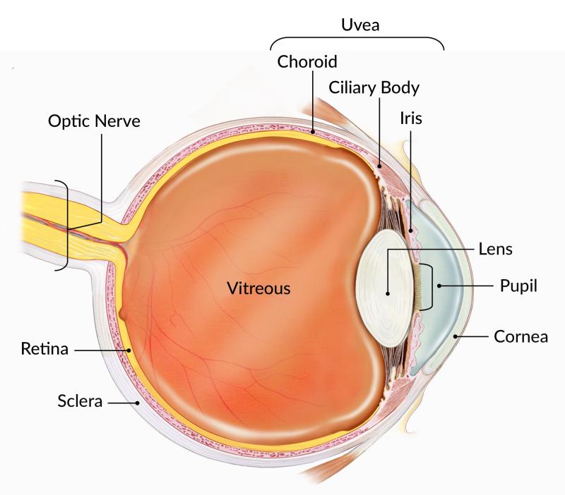
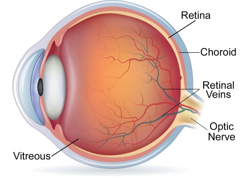
· The retina in the eyeball (diagram) (VMR Institute)
· Plan of the retinal neurons (Gray's Anatomy) (Wikimedia Commons)

Related terms
- Ciliary part of retina
- Iridial part of retina
- Non-visual retina
- Retina
- Optic disc
- Macula lutea
- Ora serrata
- Outer segment of photoreceptor
- Inner layer of eyeball
- Optic nerve
References and sources
- Further reading: Retina, Wikipedia.
- Further reading: Photoreceptor cell, Wikipedia.
- Federative International Programme for Anatomical Terminology (FIPAT). Terminologia Anatomica, 2nd ed. (TA2), 2019.