Wall of eyeball (Latin: tunicae bulbi)
Also called Coats of the eyeball
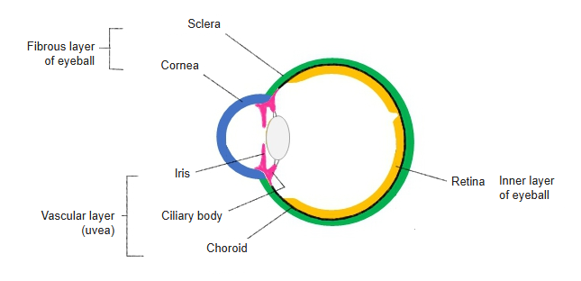
The wall of the eyeball is the set of coats that enclose the eyeball; it is made up of three layers: the outer, middle and inner layers of the eyeball.
Wall
of
eyeball
of
eyeball
- Fibrous layer : = outer layer of the eyeball.
- Cornea : the bowl-shaped front part, about one sixth of the outer layer; classically described as having five layers.
- Sclera : the thick, tough, white coat of collagen fibers that forms the rest.
- Vascular layer : = middle layer of the eyeball (uvea).
- Choroid : a thin, dark brown layer rich in blood vessels and pigment cells.
- Ciliary body : continues the choroid forwards; its main part is the ciliary muscle, which changes the thickness of the lens.
- Iris : adjusts the size of the pupil with two smooth muscles, the sphincter and the dilator pupillae.
- Inner layer : = nervous layer of the eyeball.
- Pigment epithelium : a single layer of pigment epithelium in three parts: retinal, ciliary and iridial pigment epithelium.
- Retina : two parts, the optic part and the blind part; only the optic part responds to light.
The wall surrounds the contents of the eyeball: the aqueous humor, the lens and the vitreous body. The fibrous layer gives the eye its shape and protects it; the vascular layer is rich in blood vessels and pigment. Many sources count the pigment epithelium as the outer, pigmented layer of the retina and call the whole inner layer the retina, with the light-sensitive optic part lining the back of the eye and the blind part covering the back of the ciliary body and iris.
Related terms
- Fibrous layer of eyeball
- Vascular layer of eyeball
- Inner layer of eyeball
- Eyeball
- Sclera
- Choroid
- Retina
References and sources
- Further reading: Human eye, Wikipedia.
- Federative International Programme for Anatomical Terminology (FIPAT). Terminologia Anatomica, 2nd ed. (TA2), 2019.