Subdural space (Latin: spatium subdurale)
It is found both around the brain and around the spinal cord.
It was once described as a narrow cleft between the dura and the arachnoid filled with cerebrospinal fluid. Most modern accounts treat it as a potential space that appears only when the layers are split, for example by injury, disease or loss of cerebrospinal fluid after death. In the healthy body the arachnoid lies against the inner surface of the dura, held there by the pressure of the cerebrospinal fluid in the subarachnoid space beneath it. The split runs through the innermost cell layer of the dura (the dural border cell layer) rather than between two separate membranes. Bleeding into it, usually from torn bridging veins (such as the superior cerebral veins) on their way to the dural venous sinuses, forms a subdural haematoma.
Reference websites
[Wikipedia]
· Subdural space, Wikipedia.
· Meninges, Wikipedia.
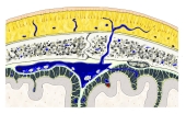 Meninges of the brain
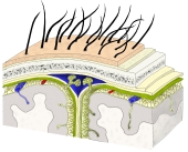 Layers from scalp to brain
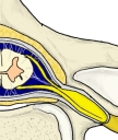 Meninges of the spinal cord
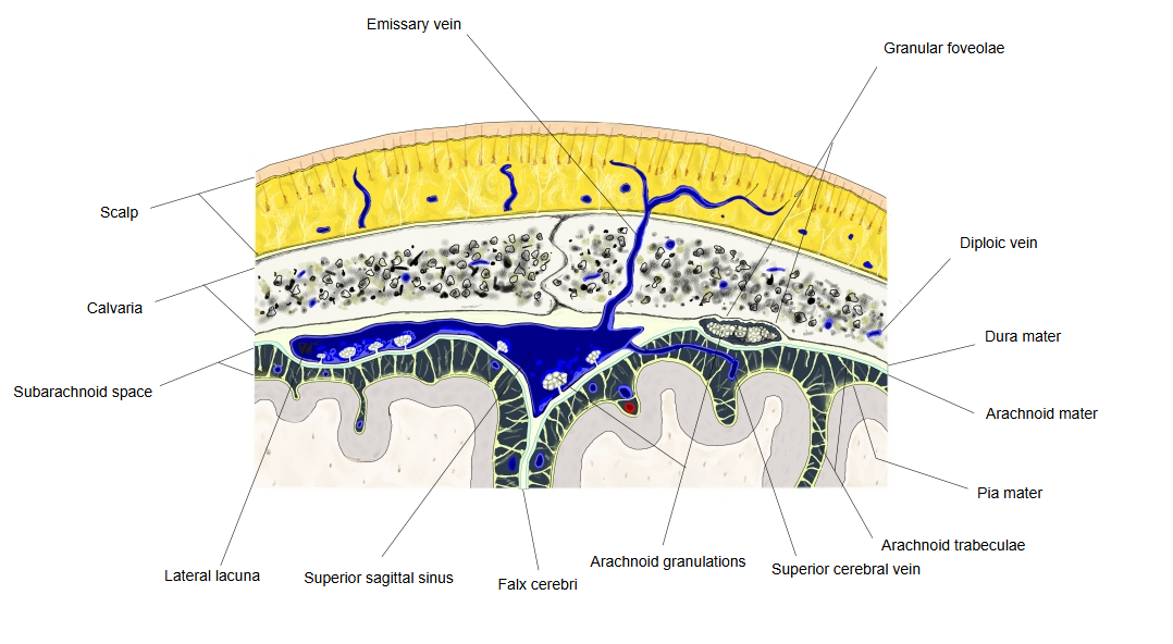
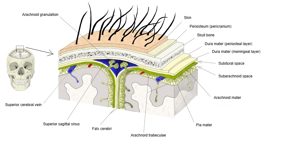
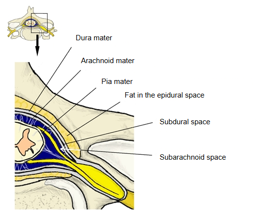
- Dura mater : outside it: the epidural space (around the spinal cord, between the spinal dura and the wall of the vertebral canal).
- Arachnoid mater : between it and the dura: the subdural space, which appears only in disease or after injury.
- Pia mater : between it and the arachnoid: the subarachnoid space, filled with cerebrospinal fluid.
Related terms
- Dura mater
- Falx cerebri
- Granular foveolae
- Dural venous sinuses
- Superior cerebral veins
- Brain
- Spinal cord
References and sources
- Further reading: Subdural space, Wikipedia.
- Further reading: Meninges, Wikipedia.
- Federative International Programme for Anatomical Terminology (FIPAT). Terminologia Anatomica, 2nd ed. (TA2), 2019.