Ciliary processes (Latin: processus ciliares)
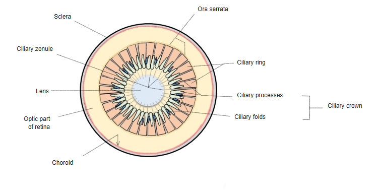
- Ciliary crown (front)
- Ciliary processes : about 70–80 radial folds rich in capillaries; they produce the aqueous humor.
- Ciliary folds : low folds between the ciliary processes.
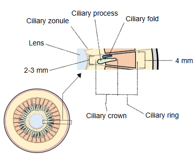
- Ciliary ring (back) : a band about 4 mm wide that meets the ora serrata; many fine ridges run on its inner surface.
· The larger processes are folds about 2–3 mm long and up to about 1 mm high; they are highest where they face the edge of the lens. Their free tips stay about 0.5 mm away from the lens.
Reference websites
[Wikipedia]
· Ciliary processes, Wikipedia.
[Websites with illustrations]
· Ciliary body and ciliary processes (figure) (Entokey)
· Schematic diagram of the human eye (Wikimedia Commons)
Note: some descriptions divide the processes into large and small ones; the small ones probably correspond to the low ciliary folds between the processes. Sources differ on whether these folds are counted as processes.
The tissue of the ciliary processes (large and small) continues from the fibrous connective tissue of the ciliary ring. As in the ciliary ring, this connective tissue framework is covered on its inner surface by the basal lamina and the ciliary part of the retina, a two-layered epithelium (an outer pigmented layer, the pigmented layer of the ciliary body, and an inner non-pigmented layer), and its outer surface meets the ciliary muscle. The processes are notably rich in vessels; their arteries come from the major arterial circle of the iris. The non-pigmented epithelium secretes the aqueous humor into the posterior chamber.
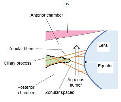
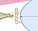 Ciliary zonule (diagram)
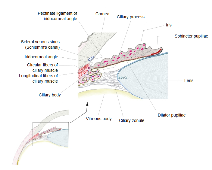
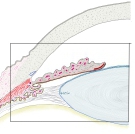 Iris and ciliary body (section)
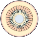 Ciliary body seen from inside
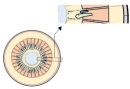 Ciliary crown and ciliary ring (enlarged)


Anatomy
The processes are arranged in a ring and form a kind of frill behind the iris, around the edge of the lens. Larger and smaller processes lie side by side; the smaller ones lie irregularly in the spaces between the larger ones. At their outer ends they are attached to ridges of the ciliary ring and are continuous with the layers of the choroid; their inner ends are free and rounded and point toward the posterior chamber and the edge of the lens. In front they are continuous with the edge of the iris (ciliary margin); behind, they are connected with the ciliary zonule (the suspensory ligament of the lens).
Function
The ciliary processes produce the aqueous humor, which fills the chambers of the eyeball.
Related terms
- Ciliary crown
- Ciliary ring
- Ciliary folds
- Ciliary zonule
- Ciliary margin of iris
- Circular fibres of ciliary muscle
- Ciliary body
- Aqueous humor
- Posterior chamber of eyeball
- Lens
- Iris
- Basal lamina of choroid
- Chambers of eyeball
References and sources
- Further reading: Ciliary processes, Wikipedia.
- Federative International Programme for Anatomical Terminology (FIPAT). Terminologia Anatomica, 2nd ed. (TA2), 2019.