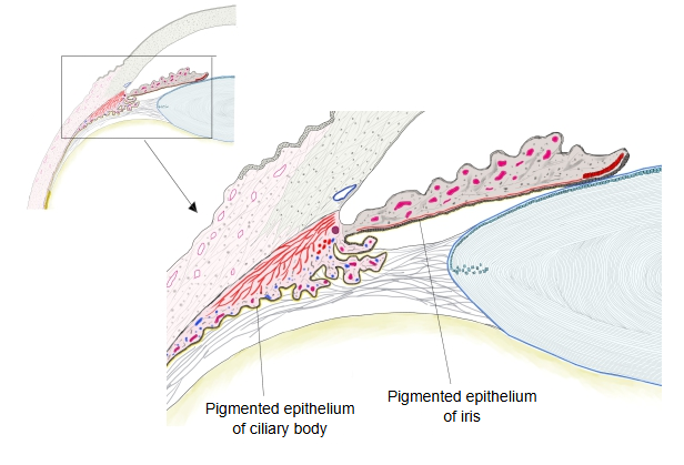
Pigmented epithelium of ciliary body (Latin: stratum pigmenti corporis ciliaris)
Also called the pigmented layer of ciliary body or the outer layer of the ciliary epithelium
surface of the
ciliary body
- Outer layer : = the pigmented epithelium of the ciliary body: a single layer of cuboidal to columnar cells rich in pigment granules (melanin).
- Inner layer : = the ciliary part of the retina in the narrow sense (in the wide sense both layers together): cuboidal to columnar cells like the outer layer, but without pigment granules; the non-pigmented epithelium.
The pigmented epithelium is the outer of the two epithelial layers that cover the inner surface of the ciliary body: a single layer of cuboidal to low columnar cells rich in melanin granules. Together with the inner, non-pigmented layer it forms the ciliary part of the retina in the wide sense, the blind continuation of the retina over the ciliary body; the two layers come from the two walls of the embryonic optic cup. At the ora serrata it continues backwards into the retinal pigment epithelium; at the root of the iris it continues forwards into the pigmented epithelium of the iris. Its cells are thinner than those of the retinal pigment epithelium and have no long microvilli. The non-pigmented layer over the ciliary processes secretes the aqueous humour.
- Retina : made up of the optic part and the blind part; only the optic part senses light, and it has many layers.
- Pigment epithelium layer : in three parts: the retinal, ciliary and iris pigment epithelia. The retinal pigment epithelium is a single layer; the ciliary and iris epithelia have two layers.
- Retinal pigment epithelium : hexagonal (prism-shaped) cells, firmly joined to each other; they send microvilli between the photoreceptor cells.
- Ciliary pigment epithelium : cuboidal cells, thinner than the retinal pigment epithelium, without microvilli.
- Iris pigment epithelium : short columnar cells covering the posterior limiting lamina of the iris from behind; at the pupillary margin the posterior layer turns forward onto the anterior layer.
Pigment epithelium of the inner layer of the eyeball, from back to front
The ora serrata is the boundary between the retinal and the ciliary pigment epithelium. The ciliary and iris pigment epithelia form the ciliary and iridial parts of the retina.
Reference websites
[Wikipedia]
· Ciliary body, Wikipedia.
[Websites with illustrations]
· Ciliary body and its epithelium (figure) (IntechOpen)

Related terms
- Ciliary body
- Ciliary part of retina
- Pigment epithelium layer
- Retinal pigment epithelium
- Pigmented epithelium of iris
- Ciliary processes
- Ora serrata
References and sources
- Further reading: Ciliary body, Wikipedia.
- Federative International Programme for Anatomical Terminology (FIPAT). Terminologia Anatomica, 2nd ed. (TA2), 2019.