Obturator internus fascia
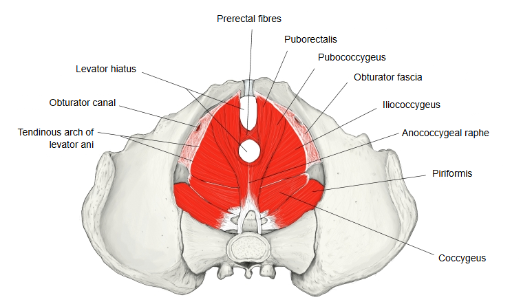
· It is a layer of the parietal pelvic fascia. Most sources call it simply the obturator fascia; some atlases use both names for the same layer.
It covers the medial (pelvic) surface of the obturator internus and is continuous with the periosteum of the bone around the origin of the muscle. It becomes stronger towards its lower part, and at the pelvic outlet it joins the falciform process of the sacrotuberous ligament. Its lower part forms the lateral wall of the ischioanal fossa, the hollow between the obturator internus and the levator ani; there it splits to enclose the internal pudendal artery and vein and the pudendal nerve in the pudendal canal. Above, at the posterior edge of the inner opening of the obturator canal, it forms a strong arched band, the tendinous arch, from which part of the levator ani arises; this band varies greatly between individuals.
See: Obturator fascia