Obturator fascia (Latin: fascia obturatoria)
Also called: obturator internus fascia
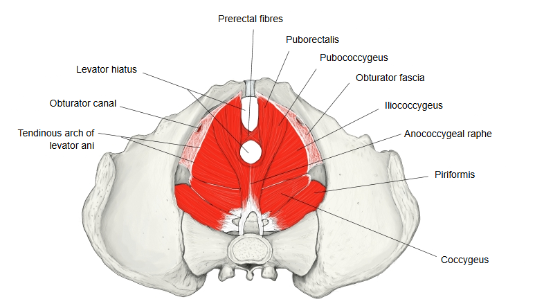
· It is part of the parietal pelvic fascia, the layer that lines the walls of the pelvis. Some textbooks list the name without describing it.
· Some atlases point to the same layer in their illustrations but call it "obturator fascia" in one place and
"obturator internus fascia" in another; the two names are used for the same layer.
It covers the pelvic (inner) surface of the obturator internus and is attached to the bone around the origin of the muscle, where it blends with the periosteum. At the top it arches below the obturator canal, leaving a gap through which the obturator vessels and nerve leave the pelvis. A thickened band within it, the tendinous arch of levator ani, runs from the back of the pubis to the ischial spine, and part of the levator ani arises from it. Below the origin of levator ani the fascia faces the perineum and forms the lateral wall of the ischio-anal fossa (the space between obturator internus and levator ani); here it splits to enclose the internal pudendal artery and vein and the pudendal nerve in the pudendal canal. It becomes stronger as it descends and, at the pelvic outlet, ends on the falciform process of the sacrotuberous ligament.
Reference websites
[Wikipedia]
· Obturator fascia, Wikipedia.
· Pudendal canal, Wikipedia.
[Websites with illustrations]
· Left hip bone with the obturator fascia and levator ani (Gray's Anatomy, fig. 404) (Wikimedia Commons)

· Tendinous arch of the pelvic fascia and of levator ani (Academic OB/GYN)

Related terms
References and sources
- Further reading: Obturator fascia, Wikipedia.
- Further reading: Pudendal canal, Wikipedia.
- Federative International Programme for Anatomical Terminology (FIPAT). Terminologia Anatomica, 2nd ed. (TA2), 2019.