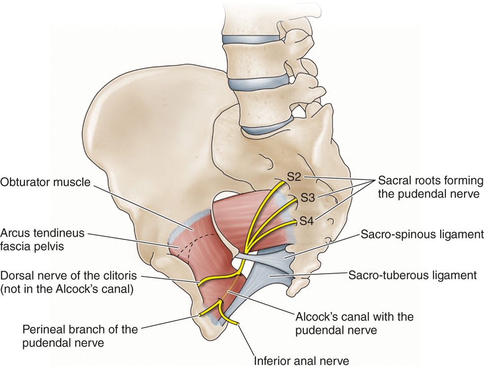
Pudendal canal (Latin: canalis pudendalis)
Also called Alcock's canal
What is the pudendal canal?
· Searching for illustrations of the canal shows two kinds of drawing:
1. 1. Seen from inside the pelvis, the canal is drawn on the lower part of obturator internus, on its inner face
(left illustration below).
2. 2. Seen from inside the pelvis, the canal is drawn at the lower edge of obturator internus, near its attachment to the ischium, wrapped in fascia
(right illustration below). Both show the same fascial sheath on the medial surface of the muscle.
passing
through
the canal
- Internal pudendal artery : one of the branches of the anterior division of the internal iliac artery (which branches from the common iliac artery).
- Internal pudendal vein : collects blood from around the anus and the external genitalia (perineum) and drains into the internal iliac vein.
- Pudendal nerve : a mixed nerve formed from the 2nd to 4th sacral nerves (spinal nerves); it supplies the skin and muscles of the perineum, including the external anal and urethral sphincters.
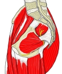 Pelvis, medial view (muscles)
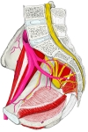 Pelvis, medial view (vessels and nerves)
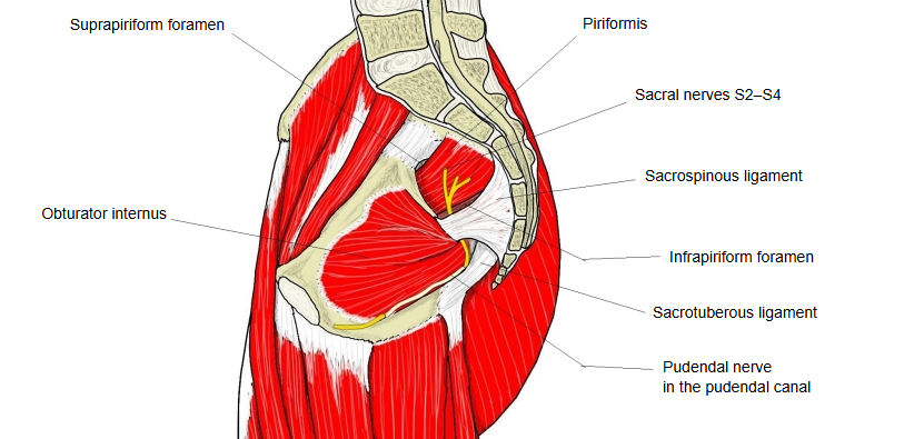
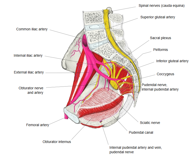
The pudendal canal (Alcock's canal) is a tunnel-like structure along the medial surface of obturator internus, formed by a split in the obturator internus fascia below the origin of levator ani. The internal pudendal artery and vein and the pudendal nerve pass through it. They leave the pelvis through the greater sciatic foramen below piriformis, hook around the sacrospinous ligament near the ischial spine, and re-enter through the lesser sciatic foramen into the canal, which runs forward to the perineum. Compression or stretching of the nerve here can cause pudendal neuralgia (perineal pain made worse by sitting).
Reference websites
[Wikipedia]
· Pudendal canal, Wikipedia.
· Pudendal nerve, Wikipedia.
[Websites with illustrations]
· Pelvic wall from inside with the internal pudendal artery and the pudendal canal (Musculoskeletal Key)

· Pudendal nerve and Alcock's canal (Anesthesia Key)
Where it is found
Related terms
- Pudendal nerve
- Internal pudendal artery
- Internal pudendal veins
- Obturator internus
- Sacrospinous ligament
- Sacrotuberous ligament
- Ischial spine
References and sources
- Further reading: Pudendal canal, Wikipedia.
- Further reading: Pudendal nerve, Wikipedia.
- Federative International Programme for Anatomical Terminology (FIPAT). Terminologia Anatomica, 2nd ed. (TA2), 2019.