Parietal pelvic fascia (Latin: fascia pelvis parietalis)
Also called the parietal fascia of the pelvis
inside the
pelvis
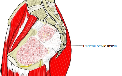
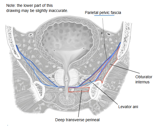
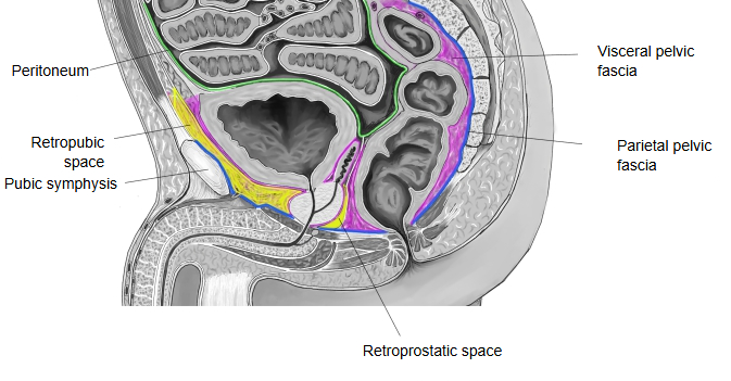
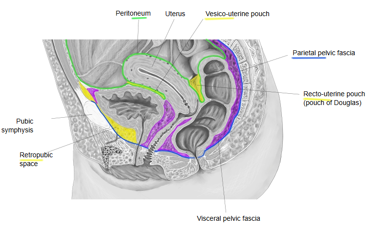
- Parietal pelvic fascia : "the fascia on the inner wall of the pelvis + the presacral fascia"; it fuses and blends with the surrounding periosteum and tendinous arches.
- Visceral pelvic fascia : covers the surfaces of the pelvic organs and fills the spaces between them (with fat and vessels, so it is thick). Its parts have their own names, such as the pubovesical ligament, the cardinal ligament and the rectovaginal septum.
· A tough membrane of collagen-rich connective tissue; it holds the vessels and nerves of the pelvis against the pelvic wall and protects them.
· It is also continuous with the visceral pelvic fascia that covers the organs.
· At the edges of these muscles it fuses with the periosteum and with tendinous arches, so it runs without a break around the pelvic wall. Above, it is continuous with the fascia lining the abdominal wall (the transversalis fascia and the iliac fascia).
The muscles on the inside of the pelvis, in brief:
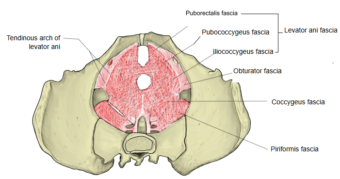
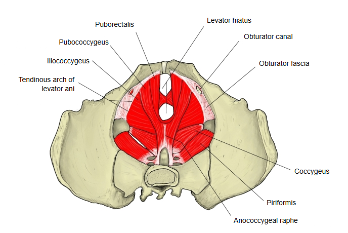
Side and back walls: obturator internus, piriformis. Floor: levator ani, coccygeus.
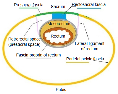
· Its parts are named after the muscles they cover: the obturator fascia, the piriformis fascia, the levator ani fascia (over the puborectalis, pubococcygeus and iliococcygeus parts) and the coccygeus fascia; in front of the sacrum it forms the presacral fascia. A thickening of the obturator fascia, the tendinous arch of levator ani, gives origin to the levator ani.
· Where the pelvic organs pass through the floor, the parietal fascia turns onto them and becomes the visceral pelvic fascia, which covers the bladder, the rectum and the uterus or prostate and fills the spaces between them with loose tissue, fat and vessels (around the rectum, the mesorectum). Spaces between the layers, such as the retropubic space and the retrorectal (presacral) space, are surgical landmarks.
Note: sources differ on the exact limits of the presacral fascia and on where the fascia ends and the periosteum begins; the boundaries drawn in the figures are approximate.
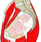 Pelvis (sagittal section)
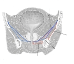 Bladder and pelvic floor (coronal section, male)
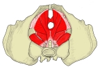 Pelvic floor muscles
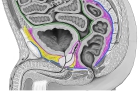 Male pelvis (median section)
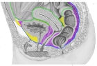 Female pelvis (median section)
Related terms
References and sources
- Further reading: Pelvic fascia, Wikipedia.
- Further reading: Endopelvic fascia, Wikipedia.
- Federative International Programme for Anatomical Terminology (FIPAT). Terminologia Anatomica, 2nd ed. (TA2), 2019.