Perichondrium (Latin: perichondrium)
Overview
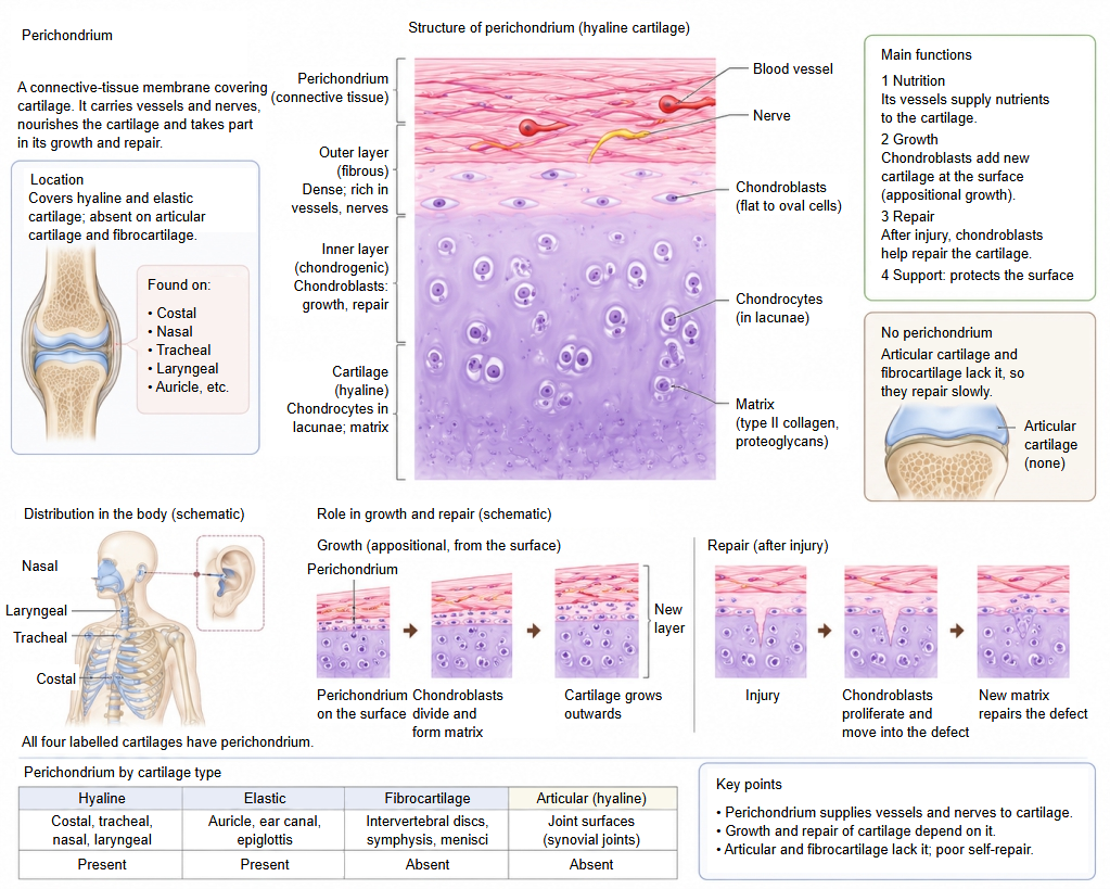
· It is usually very thin; its thickness varies with the site, with age and with the mechanical load. It covers hyaline cartilage (e.g. the costal, nasal, tracheal and laryngeal cartilages) and elastic cartilage (e.g. the auricle and the epiglottis). It is the cartilage counterpart of the periosteum of bone.
The following cartilages have no perichondrium:
1. 1. Articular cartilage (hyaline cartilage): the joint surfaces rub against each other, where nerves and vessels would be damaged; they must stay smooth and are nourished by the synovial fluid.
2. 2. Epiphyseal cartilage (hyaline cartilage): the growth plate of a child's bone, which is later replaced by bone.
3. 3. Fibrocartilage (intervertebral discs, menisci, pubic symphysis): it is rich in very strong type I collagen fibres, which merge directly with the neighbouring bone and ligaments, so it has no separate membrane.
Structure
The perichondrium has two layers.
| 1 | Outer layer (fibrous layer) | Make-up: thick bundles of mainly type I collagen (collagen fibres) and fibroblasts. Role: very strong; protects the cartilage from outside mechanical impact; rich in blood vessels and nerves. |
| 2 | Inner layer (cellular or chondrogenic layer) | Features: many chondroprogenitor cells and chondroblasts, the source of new cartilage. Role: produces new chondrocytes and matrix and makes the cartilage grow outwards from its surface (appositional growth). |
Functions
1. A supply base for nutrients and oxygen
Cartilage itself has no blood vessels at all, so nutrients and oxygen seep slowly (diffuse) into it from the vessels running in the outer layer of the perichondrium.
2. Growth, regeneration and repair of cartilage
The inner layer adds new cartilage at the surface (appositional growth). When the cartilage suffers minor damage, the cells of the inner layer become active, make new cartilage matrix and repair the injury; cartilage without perichondrium repairs poorly.
Related terms
- Cartilaginous joint
- Periosteum
- Articular cartilage
- Epiphyseal cartilage
- Costal cartilage
- Tracheal cartilages
- Laryngeal cartilages
References and sources
- Further reading: Perichondrium, Wikipedia.
- Further reading: Cartilage, Wikipedia.
- Federative International Programme for Anatomical Terminology (FIPAT). Terminologia Anatomica, 2nd ed. (TA2), 2019.