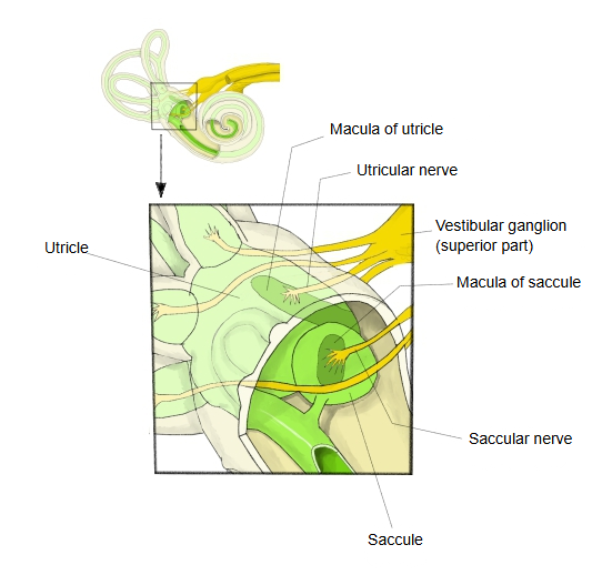
Macula of utricle (Latin: macula utriculi)
Also called Utricular macula
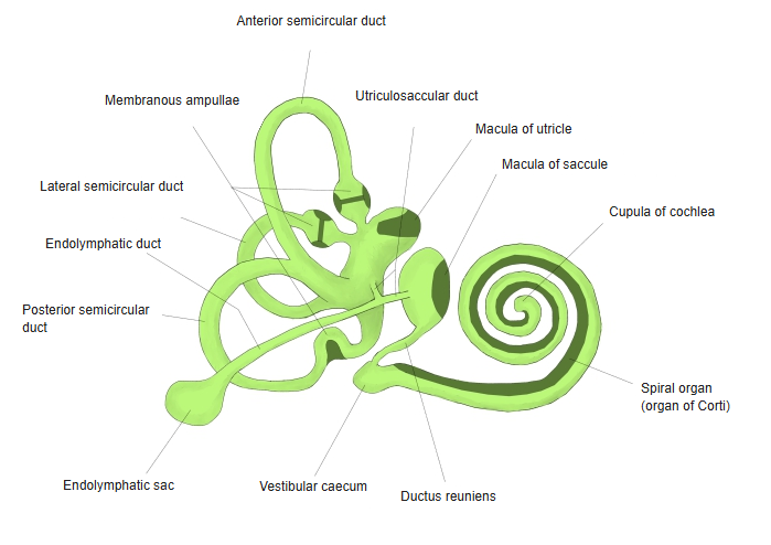
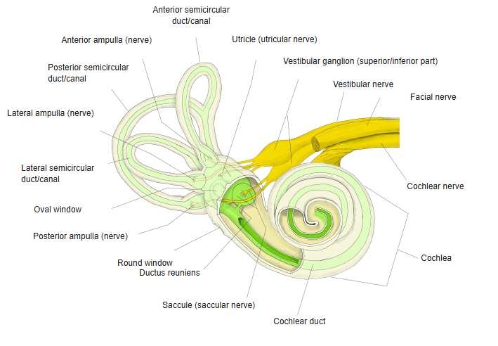
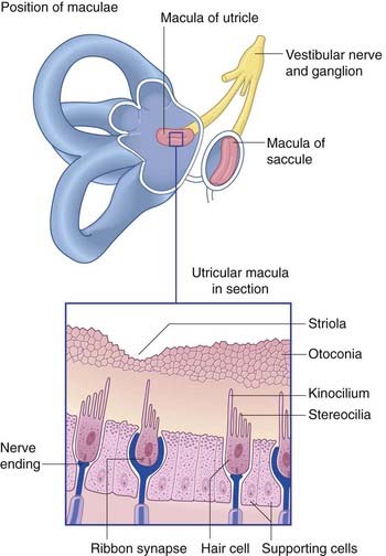
· It occupies part of the floor and part of the front wall of the utricle, and from there extends onto its lateral wall. It lies at about right angles to the macula of the saccule.
Shape: an oval, longer from side to side; about 3 mm long and about 2.4 mm wide.
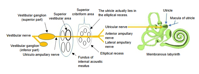
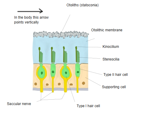
Hair cells of the maculae of the utricle and saccule
· There are two kinds of hair cells, type I and type II, with supporting cells between them.
· Each hair cell carries about 40–70 stereocilia and a single kinocilium.
· The hair bundles on top of the hair cells are covered by a jelly-like otolithic membrane.
· On the surface of the otolithic membrane lie great numbers of small crystals of calcium carbonate, the otoliths (otoconia, statoconia). When the head tilts or moves in a straight line, the heavier membrane lags behind and bends the hair bundles.
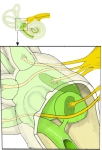 Maculae of the utricle and saccule
 Membranous labyrinth
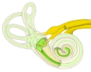 Bony and membranous labyrinth with nerves
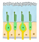 Hair cells of the macula
Reference websites
[Wikipedia]
· Macula of utricle, Wikipedia.
· Otolithic membrane, Wikipedia.
[Websites with illustrations]
· Position of the maculae and the utricular macula in section (figure) (Clinical Gate)
· How the maculae respond to tilting of the head (figure) (Pharmacy180)

Related terms
- Elliptical recess
- Utriculosaccular duct
- Vestibular maculae
- Macula of saccule
- Utricle
- Saccule
- Membranous labyrinth
- Vestibule
- Vestibular nerve
References and sources
- Further reading: Macula of utricle, Wikipedia.
- Further reading: Otolithic membrane, Wikipedia.
- Federative International Programme for Anatomical Terminology (FIPAT). Terminologia Anatomica, 2nd ed. (TA2), 2019.