Cochleariform process (Latin: processus cochleariformis)
· In the description above, the process could equally be called the end of the canal for tensor tympani, since the septum forms the floor of that canal; the meaning is the same.
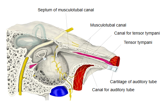
- Canal for tensor tympani : the upper of the two parts into which the septum divides the musculotubal canal; it lodges the tensor tympani.
- Septum : a thin, incomplete plate of bone that divides the musculotubal canal into an upper and a lower part; its back end curls laterally as the cochleariform process.
- Canal for auditory tube : the lower of the two parts; it forms the bony part of the auditory tube.
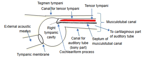
 Right tympanic cavity (sagittal section)
 Cochleariform process (oblique section)
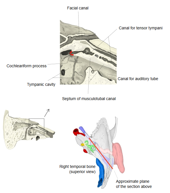
The cochleariform process is a thin bony projection on the medial wall of the tympanic cavity, towards its front, and acts as a pulley for the tendon of the tensor tympani. It lies:
at the back end of the canal for tensor tympani;
above and in front of the oval window;
just below the first part of the tympanic segment of the facial canal.
It marks the floor of the anterior epitympanic recess. The tendon of the tensor tympani leaves the canal at the tip of the process, turns sharply laterally, crosses the tympanic cavity and inserts on the medial side of the base of the handle of the malleus. The process is a useful landmark in middle ear surgery, for example for finding the facial nerve.
Reference websites
[Wikipedia]
· Tensor tympani muscle, Wikipedia.
[Websites with illustrations]
· Medial wall of the middle ear with the cochleariform process and the tensor tympani tendon (illustration) (Jaypee Digital (book figure))

· Cochleariform process and tensor tympani muscle (illustration) (Stanford Otologic Surgery Atlas)

· Lateral wall of the tympanic cavity with the cochleariform process (Sobotta, 1909) (Wikimedia Commons)

Related terms
- Canal for tensor tympani
- Septum of musculotubal canal
- Musculotubal canal
- Tensor tympani
- Tympanic cavity
- Malleus
- Oval window
References and sources
- Further reading: Tensor tympani muscle, Wikipedia.
- Federative International Programme for Anatomical Terminology (FIPAT). Terminologia Anatomica, 2nd ed. (TA2), 2019.