Anterior limiting lamina of iris
Also called the anterior border layer of iris
· It is made of fairly dense connective tissue mixed with pigment cells.
The colour of the iris is said to depend on the amount of pigment in this layer.
It faces the anterior chamber and is made of closely packed fibroblasts and melanocytes. The amount of pigment in this layer and in the stroma is a main factor in eye colour: little pigment gives a blue eye, much pigment a brown eye. The layer is incomplete in places, leaving pits (crypts) through which the stroma is open to the aqueous humor of the anterior chamber.
· A layer with the same name is found in the cornea: the anterior limiting lamina of the cornea (Bowman's layer).
of the
iris
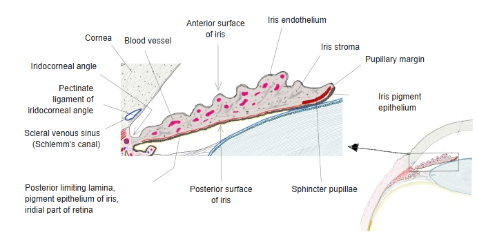
- Iris endothelium : described as a layer of flattened cells continuing the corneal endothelium (the endothelium of the anterior chamber); in the adult human iris it is not a continuous layer
- Stroma of iris
- Anterior limiting lamina : fairly dense connective tissue with pigment cells
- Vascular layer : the main part of the iris, rich in vessels and nerves; it contains the sphincter pupillae (the dilator pupillae lies at its back)
- Posterior limiting lamina
- Behind it : the pigmented epithelium of the iris, which belongs to the inner layer of the eyeball (the iridial part of the retina)
The table above follows one source; the layers of the iris are described differently in different sources.
Wikipedia, for example, divides the iris broadly into two layers: the stroma and the pigmented epithelial cells.
Related terms
- Anterior pole of eyeball
- Anterior chamber of eyeball
- Iris
- Posterior limiting lamina of iris
- Folds of iris
- Pupil
- Chambers of eyeball
- Vascular layer of eyeball
References and sources
- Further reading: Iris (anatomy), Wikipedia.
- Federative International Programme for Anatomical Terminology (FIPAT). Terminologia Anatomica, 2nd ed. (TA2), 2019.