Superior medullary velum (Latin: velum medullare superius)
Also called the anterior medullary velum
In more detail:
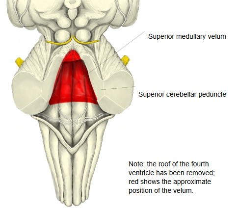
The superior medullary velum is a thin plate of white matter between the two superior cerebellar peduncles that forms the roof of the upper part of the fourth ventricle. Its back (dorsal) surface is fused with the lingula of the cerebellum.
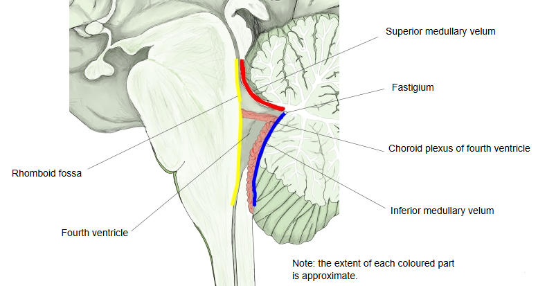
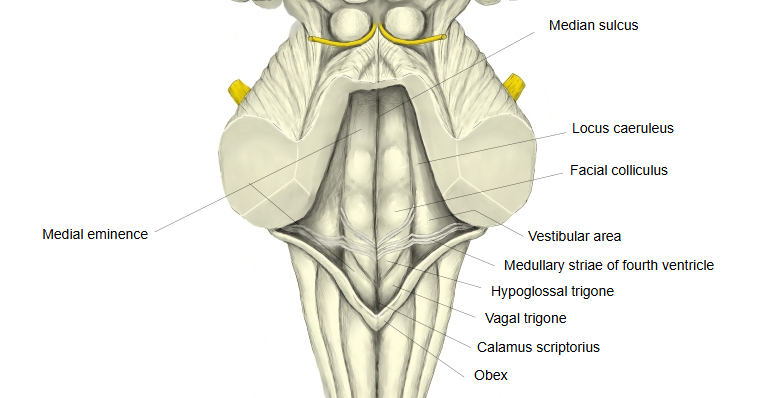
The roof of the fourth ventricle is tent-shaped: the superior cerebellar peduncles and the superior medullary velum form the upper slope, and the inferior medullary velum and the tela choroidea form the lower slope. The floor is the rhomboid fossa.
The velum is narrow above, below the inferior colliculi of the midbrain, and widens below, where it is continuous with the white matter of the cerebellar vermis. The trochlear nerves emerge from the back of the midbrain just above it, and their fibres cross (decussate) inside the velum. It is supplied by branches of the superior cerebellar artery.
Some textbooks divide the roof of the fourth ventricle into a front (upper) half and a back (lower) half, made up as follows:
fourth
ventricle
- Upper (front) half : the superior medullary velum and the superior cerebellar peduncles (brachia conjunctiva).
- Lower (back) half : the inferior medullary velum and the choroid plexus (tela choroidea) of the fourth ventricle.
Note: some sources also count the nodule of the cerebellar vermis as part of the lower half.
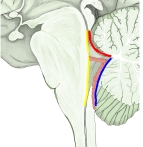 Fourth ventricle (median section)
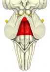 Brainstem, posterior view (cerebellum removed)
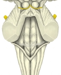 Floor of fourth ventricle
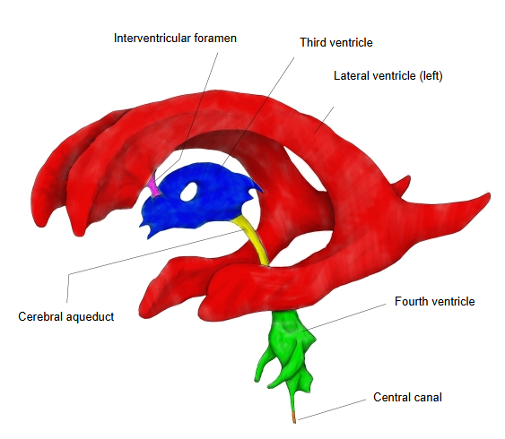
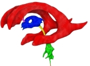 Ventricles: left side (coloured)
Reference websites
[Wikipedia]
· Superior medullary velum, Wikipedia.
· Fourth ventricle, Wikipedia.
Related terms
- Inferior medullary velum
- Rhomboid fossa
- Cerebral ventricles
- Third ventricle
- Superior cerebellar artery
- Trochlear nerve
- Brain
References and sources
- Further reading: Superior medullary velum, Wikipedia.
- Further reading: Fourth ventricle, Wikipedia.
- Federative International Programme for Anatomical Terminology (FIPAT). Terminologia Anatomica, 2nd ed. (TA2), 2019.